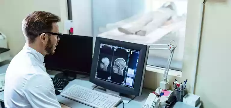
Medical diagnostics is an ever-evolving landscape in which there is enough significance in brain imaging. With its ability to go deeper into the intricate complexities of the human brain MRI (Magnetic Resonance Imaging) has...
Introduction
Medical diagnostics is an ever-evolving landscape in which there is enough significance in brain imaging. With its ability to go deeper into the intricate complexities of the human brain MRI (Magnetic Resonance Imaging) has provided a newer understanding of neurological disorders and catapulted patient care to great heights.
Among a bevy of amazing imaging modalities, MRI has great importance as a potent tool for visualizing the brain in exquisite detail. Based on the principles of magnetic fields and radio waves, MRI offers unparalleled insights into the structure and function of the brain, enabling doctors to unravel many mysteries wrapped within its delicate folds.
In this article, we will walk through the remarkable capabilities of an MRI that assist in the treatment of the brain. We’ll discover how it has contributed to the advancements in medical diagnostics and patient care.
A few basics about MRI
There is some fantastic interplay of basic principles in an MRI process that facilitates viewing the brain's intricate structures. MRI technology operates based on the premise of magnetism, radio waves, and image formation.
The MRI scanner is capable of generating a powerful magnetic field, which aligns the hydrogen atoms in the body's tissues. Once radio waves are applied, these aligned hydrogen atoms begin to emit signals that are detected by specialized coils within the scanner. By utilizing the timing and strength of the radio waves, the MRI system can encode spatial information. After that, there is processing and transformation of this information translating into detailed cross-sectional images of the brain. An MRI displays various tissue types and highlights abnormalities.
So, the components of an MRI scanner include the magnet, gradient coils, radiofrequency coils, and computer systems. All these components work in tandem to generate high-resolution images offering healthcare professionals invaluable insights into the brain's anatomy and function. Thus, Brain MRI has carved a path leading to precise diagnoses and optimized treatment strategies for patients.
Different types of Brain MRI
There are a slew of different MRIs for diagnosing and treating the human brain. Each presents its specific advantages in helping in the intervention of brain diseases.
Structural MRI
Structural MRI provides valuable insights into various neurological conditions, where T1-weighted images can provide excellent anatomical detail, highlighting different brain structures. It is helpful in the detection of lesions or abnormalities.
On the other hand, T2-weighted imaging is focused on illuminating the differences in tissue properties, helping in identifying edema, inflammation, or tumors.
Another technique called fluid-attenuated inversion recovery (FLAIR), selectively suppresses the signal from cerebrospinal fluid, bettering the visibility of lesions, such as multiple sclerosis plaques.
Gradient echo (GRE) is another technique, which is particularly useful in detecting hemorrhages, microbleeds, or certain vascular malformations. All these imaging sequences help doctors assess brain morphology, identify pathological changes, and make treatment decisions.
Functional MRI
Functional MRI (fMRI) as a powerful imaging tool provides great insights into the dynamic functioning of the human brain. It hovers around the principles of blood oxygenation level-dependent (BOLD) contrast, which is based on the interplay between neural activity, cerebral blood flow, and oxygenation. By monitoring and detecting these changes, fMRI maps brain activity and spots regions involved in specific tasks or at rest. fMRI has contributed immensely to the research of cognition, emotion, language, and sensory processing, throwing light on brain function and its disruptions in various neurological conditions.
Diffusion-weighted imaging (DWI)
It is an MRI technique that provides unique insights into the movement of water molecules within brain tissue. By measuring the diffusion of water molecules, DWI can provide important information about the microstructure and integrity of white matter tracts in the brain. It helps doctors in detecting changes in diffusion patterns, helping in the diagnosis and monitoring of conditions such as multiple sclerosis, traumatic brain injury, and neurodegenerative disorders.
Apart, DWI also helps in the early detection and characterization of acute stroke by identifying areas of restricted diffusion, signaling ischemic tissue damage. This makes DWI an important tool in both research and clinical practice, enabling healthcare professionals to have a better understanding of conditions affecting brain integrity and acute cerebrovascular events.
Perfusion-weighted imaging (PWI)
Perfusion-weighted imaging (PWI) provides information on cerebral blood flow and perfusion abnormalities within the brain. PWI is a great imaging technique helping in the assessment of regional blood flow while identifying areas with impaired perfusion. PWI has widespread clinical applications, especially in the evaluation of acute stroke, where it can help identify areas of reduced blood flow, guide treatment decisions, and assess the effectiveness of interventions.
PWI can provide information about tumor vascularity and help differentiate between tumor types. It can also assist in diagnosing and managing vascular disorders such as arteriovenous malformations and vasculitis, facilitating clinicians to assess perfusion deficits and plan appropriate interventions.
Advanced Brain MRI Techniques
Advanced techniques in brain MRI such as Magnetic resonance spectroscopy (MRS), susceptibility-weighted imaging (SWI), and Diffusion tensor imaging (DTI) offer great assistance to the healthcare professionals involved in the treatment of brain diseases.
MRS is a non-invasive technique helping in the assessment of brain metabolism and chemical composition. By measuring specific metabolites, MRS provides valuable insights into various neurological conditions, including tumor grading, neurodegenerative diseases, and epilepsy. It is a great contribution to diagnosing and planning treatment interventions.
DTI, on the other hand, measures the microstructure and connectivity of white matter in the brain. It improves the visualization of neural pathways through tractography, shedding light on brain connectivity and integrity. DTI also helps in understanding brain development, mapping fiber tracts, and studying conditions such as traumatic brain injury and neurodegenerative disorders.
SWI boosts the detection of microhemorrhages, iron deposition, and venous structures in the brain. It has a significant contribution to the evaluation of neurovascular diseases and neurodegeneration, offering insights into pathologies that may not be easily accessible with conventional MRI sequences.
Brain MRI price
Are you concerned about the cost of a brain MRI? Brain MRI prices can vary based on several factors. It can be influenced by factors like the healthcare facility, location of the facility, and specific requirements of the scan.
Other factors that govern the overall cost may include the type of MRI scan (structural, functional, etc.), the need for contrast agents, and any other sequences or specialized techniques. You should also know that these prices are independent of other related expenses such as consultation fees, radiologist interpretation fees, or any follow-up tests or procedures that may be necessary.
Your insurance coverage and individual healthcare plans also have a say in determining the out-of-pocket costs. So, you should talk to your healthcare providers and insurance companies to get a fair idea about the pricing and coverage details for brain MRI scans.
Conclusion
Hence, we now know that brain MRI is a powerful and non-invasive imaging technique helping healthcare professionals to procure detailed images of the brain's structure and function. By capitalizing magnetic fields and radio waves an MRI of the brain can provide valuable insights into various neurological conditions. It is a boon for patients and doctors alike in the diagnosis, treatment planning, and monitoring of brain diseases.
This is a painless procedure that helps in studying abnormalities, such as tumors, strokes, and neurodegenerative diseases. It empowers doctors to make informed decisions and provide personalized care to their patients. They can capture high-resolution images of the brain, and continue to be an invaluable tool in neuroscience. There are also perpetual advancements in this field, enhancing the understanding of the human brain and bettering patient outcomes.
FAQs
What are the factors a brain MRI price typically depends on?
The price of a brain MRI can vary depending on factors such as location, healthcare facility, and specific requirements of the scan.
Does my insurance cover the cost of a brain MRI?
Insurance coverage for a brain MRI is largely dependent on the patient’s insurance plan. You must contact the insurance provider to understand the coverage details, including deductibles, co-pays, and any pre-authorization requirements.
Are there additional costs associated with a brain MRI?
In addition to the MRI itself, there may be additional costs such as consultation fees, radiologist interpretation fees, or the use of contrast agents if required. It's essential to inquire about these potential additional expenses.
Can the cost of a brain MRI differ between different healthcare facilities?
Yes, the cost of a brain MRI can vary between healthcare centers. Factors such as location, facility reputation, and quality of equipment contribute to the price difference. So, try to do some price comparisons and check the quality of services.


