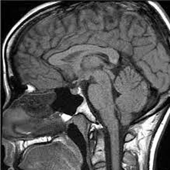
Pituitary gland is a small oval shaped endocrine gland of our body, also known as Master gland as it regulates the function of other glands of our body. It secretes various hormones into the bloodstream. It has two lobes,...
What is MRI Pituitary Gland?
Pituitary gland is a small oval shaped endocrine gland of our body, also known as Master gland as it regulates the function of other glands of our body. It secretes various hormones into the bloodstream. It has two lobes, namely the anterior lobe and the posterior lobe.
Hormones secreted by the anterior lobe includes
1) TSH (Thyroid stimulating hormone)
2) Prolactin
3) ACTH (Adrenocorticotropic hormone)
4) Growth hormone
5) FSH (Follicle stimulating hormone)
6) LH (Luteinizing hormone)
7) Melanocyte stimulating hormone
8) Enkephalins
9) Endorphins
Hormones secreted by the posterior lobe of pituitary gland include:
1) Oxytocin
2) ADH also known as Vasopressin
MRI Pituitary gland is a collection of MRI sequences put together to increase sensitivity and specificity for the evaluation of lesions of pituitary gland such as pituitary adenoma and various other sellar and suprasellar anomalies.
Now-a-days, MRI is considered as the imaging modality of choice for pituitary gland evaluation.
What are the uses of MRI Pituitary Gland?
MRI pituitary gland help in the assessment and evaluation of:
* Pituitary gland including infundibulum and posterior pituitary bright spot and the adjacent areas.
* Territory of cavernous sinus and Meckel’s cave.
* Optic nerves , optic chiasma and optic tracts.
* Internal carotid arteries and its branches.
* Diaphragma sellae and the boundaries of pituitary fossa.
MRI pituitary gland is indicated for the identification and assessment of the following:
1) Pituitary masses including solid mass, cystic mass or mixed cystic and solid pituitary mass.
2) Intrasellar, suprasellar or both supra and intrasellar masses.
3) Erosion of sellae, changes in size of sella.
4) Displacement of diaphragma sella.
5) Carotid narrowing.
6) Dural tail due to hypophysitis, meningioma aur metastases.
7) Invasion of cavernous sinus, sphenoid sinus or orbit by tumors.
8) Aneurysms or any other vascular abnormalities located especially in the cavernous sinuses.
9) Aberration of carotid arteries or other vessels.
10) Sphenoid septum, extent of pneumatization of sphenoid sinus or any anomaly in the anatomy of sinus.
11) Any bony dehiscence in the sphenoid sinus.
12) Compression of optic nerve, optic chiasma and optic tracts.
13) Pituitary adenoma, craniopharyngioma, meningioma or any metastases.
14) Ectopic posterior pituitary.
15) Sheehan syndrome or ischemic necrosis of Pituitary gland.
What are the patient preparations done for MRI Pituitary Gland?
You may follow these steps before going for MRI Pituitary gland:
1) Fix an appointment- Schedule your appointment in a Diagnostic center having the facility of MRI Pituitary gland.
2) Food and medications- follow your daily diet and usual medications unless specified otherwise by your Health care professional.
3) Clothing- Dress yourself in loose and comfortable outfits that are easy to put on and off as you need to change your clothes before MRI.
4) Pregnancy- inform your doctor about pregnancy if you are pregnant as certain medications are avoided in pregnancy for the safety of your baby. Your doctor need to assess the benefit and the risks associated.
5) Implants- Any metallic implants need to be informed, as MRI uses magnetic field to obtain images and this magnetic field can cause health hazards and safety issues if you have any metallic or electronic implants in your body such as cochlear implants, cardiac implants, metallic dental braces etc.
6) Anxiety or Claustrophobia- If you have anxiety problems or in case you are claustrophobic then tell your Doctor about these. He may prescribe sedatives or some other alternatives to help you relax while doing the scan.
7) Allery- Prior history of allergy to any medications or drugs should be informed before MRI scan.
8) Medical reports- Carry your relevant medical and lab reports along with you while going for MRI Pituitary gland. These reports may help your doctor to reach at the diagnosis.
9) Go with friend or family member to have mental and emotional support or to drive you home back.
What is the procedure for MRI Pituitary Gland?
Procedure of MRI Pituitary gland may include the following steps:
1) Before entering the MRI scanner room , you have to give a written consent.
2) You will be then asked to remove all metallic elements such as jewellary, bra with metallic underwiring, metallic hair clips, wallets, cards containing metallic strips, belts, coins, goggles, belts, hearing aids etc.
3) If you have anxiety disorder or claustrophobia then you may be given sedatives or some other alternatives to help you relax during the scan.
4) MRI machine makes banging sounds and to overcome this ,you may be provided with ear plugs or head phones to help you feel more relax and comfortable and shield you from the annoying noise produced by the MRI machine.
5) You will get thoroughly instructed before the scan begin.
6) After that you will be asked to take off your clothes and wear a gown provided to you by the technician assisting the procedure .
7) If contrast MRI is indicated then your assisting Doctor will address about all the possible side effects of using contrast agent such as rash, swelling , itching etc. Also, your Healthcare provider may check your KFT report to rule out any Renal pathology/disease and to check your GFR( Glomerular filtration rate). As contrast agent, Gadolinium should not be given to a patient having a GFR <30.
8) Then you will be positioned on MRI table which will slide and place you inside the MRI scanner.
9) You need to remain very still during the scan as body movements may interfere with the quality of images produced.
10) Your Radiologist will take several images of the pituitary gland and the adjacent structures to evaluate the underlying pathology and at the end ,these images will be interpretated for making a definite diagnosis.
11) Once the procedure gets over you may be allowed to exit the MRI scanner room. However, If contrast MRI Pituitary is being done then you have to sit in the observation room for sometime, after the scan get done , in order to check for any possible adverse effect of contrast agent being used.
12) You will get reports on the next day. However, you may get the image films on the same day itself if required by your Doctor.
How much time taken to do MRI Pituitary Gland?
MRI uses magnetic field to create detailed images and delineate the anatomy of Pituitary gland and the associated structures that help in the diagnosis and evaluation of any pathological condition of pituitary gland or the adjacent structures.
A targeted MRI scan of the pituitary gland and the adjacent region includes sagittal and coronal view, T1 weighted post-contrast pictures , and dynamic contrast-enhanced coronal images, which are essential for the detection of small pituitary microadenomas. T2 weighted images are also obtained , although are of relatively less significance.
It is a non-invasive procedure and usually takes about 30-60 minutes.
However, it may last longer too depending upon the severity of condition and associated co-morbidities or if contrast agents are being used during the procedure..
What are the charges of MRI Pituitary Gland?
Cost of MRI Pituitary gland may vary with city, locality and from center to center depending upon the quality of service and offers availing in them.
Ganesh Diagnostic and Imaging Center is offering MRI Pituitary gland at 50% Discount. The actual price of MRI Pituitary gland is Rs 8000 and you can avail this test at just RS 4000.
MRI Pituitary Gland
Rs 800 - Rs 400 Book Now
MRI Brain Of Pituitary
Rs 800 - Rs 400 Book Now
MRI Brain With Pituitary
Rs 800 - Rs 400 Book Now
MRI Pituitary
Rs 800 - Rs 400 Book Now
MRI Pituitary With Contrast
Rs 800 - Rs 400 Book Now
MRI Pituitary Gland
Rs 800 - Rs 400 Book Now
Ganesh Diagnostic and Imaging Center is a NABH accredited top diagnostic center situated in ROHINI and various other locations of Delhi. It is equipped with modern ,highly expensive machines with latest cutting edge technologies and highly skilled Radiologists and Pathologists.
We are open 24*7 and 365 day. You can also get free Consultation with our Senior Radiologist, Dr. Ravin Sharma regarding any imaging and test procedure.
We also offer facilities of online reporting, free home sample collection and free Ambulance services in Delhi, NCR. We are also empanelled with various departments and organizations.So you can get the services at panel rate too.
Grab the best deals now!


