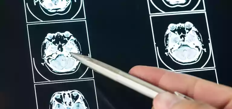
An MRI (magnetic resonance imaging) stroke protocol is referred to a specific set of imaging techniques and sequences that help to evaluate and diagnose strokes using magnetic resonance imaging.
Introduction
An MRI (magnetic resonance imaging) stroke protocol is referred to a specific set of imaging techniques and sequences that help to evaluate and diagnose strokes using magnetic resonance imaging. The main purpose of this protocol is to capture detailed and accurate images of the brain, helping doctors to identify the presence, location, and extent of a stroke.
A few key imaging sequences of MRI stroke protocol
Some of the prominent imaging sequences of MRI stroke protocol include:
Diffusion-weighted imaging (DWI): This is very sensitive for acute ischemic strokes. DWI is capable of measuring water molecule movement in the brain. It pinpoints the restricted diffusion areas, which indicates a stroke.
Fluid-attenuated inversion recovery (FLAIR): In this imaging sequence, it is possible to view brain tissue and it suppresses cerebrospinal fluid (CSF) signals. It helps in identifying swelling (edema) and detects the extent of the stroke.
T1-weighted imaging: This provides anatomical information about the brain, and helps in viewing structural abnormalities like tumors or vascular malformations that can be similar to stroke symptoms.
T2-weighted imaging: T2-weighted images are sensitive to changes in tissue water content and help in detecting brain injury areas and edema.
Magnetic resonance angiography (MRA): It evaluates blood vessels in the brain and neck, handing information about artery patency, stenosis (narrowing), or occlusion. This is also helpful in identifying the underlying cause of the stroke, such as a blood clot or aneurysm.
By binding these imaging sequences together, the MRI stroke protocol empowers doctors in finding out the location, size, and type of stroke, whether it is ischemic or hemorrhagic. Apart from this, it helps to differentiate a stroke from other conditions that may have similar symptoms. This crucial knowledge from the imaging sequences guides treatment decisions and helps in ascertaining the correct management and treatment interventions for stroke patients.
Importance of MRI Stroke Protocol
As we now know that the MRI Stroke Protocol is an important imaging technique with a host of benefits in stroke diagnosis and treatment. This is an advanced protocol, which helps doctors to procure high-resolution images of the brain, providing crucial information about the extent and location of stroke-related damage.
An MRI Stroke Protocol is known for its swift image-capturing ability of brain structures, blood vessels, and abnormalities, aiding physicians to make accurate and timely diagnoses, so that they can devise appropriate treatment plans.
On top of it, it has considerable advantages over other imaging techniques, such as computed tomography (CT) because it is non-invasive, radiation-free, and capable of providing superior soft tissue contrast.
With its quality to identify ischemic or hemorrhagic strokes, determine the viability of brain tissue, and monitor the effectiveness of treatment interventions, the MRI Stroke Protocol is an indispensable tool in guiding clinical decisions, optimizing patient welfare, and increasing our understanding of stroke pathology.
Challenges and clinical applications
The MRI Stroke Protocol has a wide range of clinical applications in the management of acute stroke, bringing about a paradigm shift in the ways how strokes are diagnosed and treated. Its capability to click precise high-resolution images of the brain assists doctors in accurately assessing the extent and location of stroke-related damage.
It can identify the type of stroke, give a view of the affected blood vessels, and assess the viability of brain tissue. Moreover, it also helps in guiding interventions such as thrombolysis or endovascular clot retrieval, making sure that the patient receives timely and effective care.
Yet, there are certain limitations while implementing MRI Stroke Protocol. One of the topmost challenges is the time constraints and urgency associated with stroke cases. In the case of managing acute stroke, time is crucial and requires prompt action. But, an MRI may not be immediately accessible or possible. It has limited availability, there are scheduling issues, or shortage of specialized staff in real-time. The decisions that are needed to be taken in such a situation are time-sensitive, but it is difficult to take them usually. So, despite having potential benefits, an MRI may fail to deal with the urgency of treatment.
Another drawback is the patient eligibility and contraindications, in capitalizing on the MRI Stroke Protocol. Some patients, such as those with pacemakers, metallic implants, or severe claustrophobia, may not qualify for MRI scans. In such cases, alternative imaging techniques like CT scans may be preferred by the doctor.
Moreover, patients who are unstable or are unable to lie still for an extended period may have difficulty undergoing MRI, which is again another drawback.
On the other hand, another important consideration is the availability and resources for MRI facilities. It is especially true in rural or underserved areas. The high cost and maintenance requirements of MRI machines may limit their accessibility, posing a threat and challenge to stroke care between different regions. Again, to have equitable access to MRI Stroke Protocol calls for great investments in infrastructure, training healthcare providers, and creating referral networks to expedite timely and appropriate imaging for stroke patients.
Yet, despite these challenges, there is no denying the fact that MRI Stroke Protocol is a valuable tool in acute stroke management. Addressing the limitations and considering the specific circumstance of each patient can augment its clinical utility and contribute to improved outcomes for individuals who suffer a stroke.
With the advancement of technology and modern science, MRI stroke protocol has also made huge strides toward more evolved techniques. In the coming days, it is surely going to address the current shortfalls and help in adequately providing treatment solutions to stroke patients.
MRI stroke protocol price
Since this is an advanced diagnostic tool, the MRI stroke protocol price is going to be high, which is a concern for certain patients. However, one should realize that the price of an MRI stroke protocol may vary based on numerous factors. It depends on the neighborhood of the facility, how advanced and modern is the facility, the use of contrast agents, the need for any extra screenings, etc. However, despite the high MRI stroke protocol price, one should not skip this important diagnostic tool if ordered by the doctor. It will compromise the safety and health of the patient to evade this scan. So, one should directly talk to the facility, and the insurance company and investigate if there are any special discounted prices available in any healthcare facility.
Conclusion
So, we have seen that the MRI stroke protocol has great importance in stroke management. With its ability to provide detailed and accurate images, doctors can make quick and informed decisions regarding stroke diagnosis and treatment. It indeed has certain challenges too. Yet, despite these challenges such as time constraints and patient eligibility, it plays a vital role in improving patient outcomes.
Even in times ahead, continued advancements in MRI technology and increased accessibility to MRI facilities make this tool promising to enhance stroke management, leading to better prognoses and improved quality of life for stroke patients.
FAQs
Is an MRI considered a part of stroke protocol?
Yes, an MRI (Magnetic Resonance Imaging) is indeed considered a crucial component of the stroke protocol. The MRI Stroke Protocol refers to a specific set of imaging techniques and sequences used to obtain detailed images of the brain in stroke cases.


