The bacterium Rickettsia akari is the source of the infectious illness rickettsialpox. It is transferred to people when a mouse mite, usually a common domestic mouse mite, bites them.
The bacterium Rickettsia akari is the source of the infectious illness rickettsialpox. It is transferred to people when a mouse mite, usually a common domestic mouse mite, bites them.
Typically, rickettsialpox is a self-limited condition that goes away on its own in two to three weeks, however, medications might hasten o heal and lessen the intensity of symptoms. In serious situations, hospitalization can be necessary.
Insect repellents, wearing protective gear when working in locations with a high risk of exposure, and keeping homes and workplaces clean and free of rodent infestations are all ways to prevent contact with mouse mites and prevent the spread of rickettsialpox.
This article will enlighten you about rickettsialpox in-depth.
From Discovery to Diagnosis: The Evolution of Rickettsialpox
When an outbreak struck tenement dwellers in New York City in 1946, rickettsialpox was first identified as a unique illness. Due to the way the rash looked, the illness was originally misdiagnosed as chickenpox, but laboratory tests revealed the presence of a rickettsial agent that had never been seen before.
Further research established that the epidemic was most likely brought on by mouse mites that had invaded the structure and were biting people. Studies revealed that the bacteria that caused the illness was closely linked to the one that causes epidemic typhus, Rickettsia prowazekii.
Although isolated instances continue to be documented in metropolitan areas with high rat infestation levels, the disease is often thought to be uncommon.
Rickettsialpox: A Closer Look at the Bacteria that Cause the Illness
The causative agent of rickettsialpox is a bacterium called Rickettsia akari.
A tiny, pleomorphic bacterium called Rickettsia akari has a range of sizes and shapes depending on its life cycle stage and the host cell it is living in.
Rickettsia akari manifests as a coccobacillus, a small, rod-shaped bacterium with a slightly rounded end, in its extracellular form.
Rickettsia akari appears pleomorphic or polymorphic in its intracellular form, with irregular cell forms that range from cocci (spherical cells) to bacilli (rod-shaped cells).
As an obligate intracellular pathogen, Rickettsia akari is dependent on the presence of host cells to reproduce and spread illness.
Rickettsia akari, like other rickettsial infections, has an unusual cell wall structure that excludes peptidoglycan, a crucial element of bacterial cell walls. Instead, Rickettsia Akari's cell wall is made up of proteins and lipopolysaccharides that aid the bacteria in thwarting host immune responses.
Using electron microscopy, it is possible to see aggregates or clusters that Rickettsia akari may develop inside host cells. These collections, often referred to as microcolonies, may have had an impact on the development of rickettsialpox.
Rickettsialpox: Recognizing the Symptoms of a Flea-Borne Infection
The symptoms of Rickettsia akari infection, often known as rickettsialpox, include:
After being bitten by an infected mite, rickettsialpox normally takes 5 to 10 days to incubate.
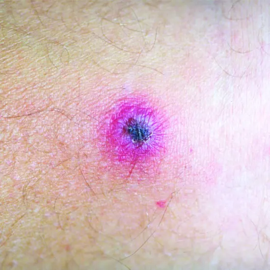
The emergence of a tiny, painless, red papule at the site of the mite bite is the first sign of rickettsialpox. This papule frequently itches and may seem like a flea bite or a pimple.
The papule turns into a vesicle (a little blister) after 1-2 days. From there, it could become a scab or crust.
Patients may experience systemic symptoms of rickettsialpox after the papule appears, such as fever, headache, muscular pains, and weariness. A generalized rash that generally covers the trunk, arms, and legs may appear in addition to these symptoms.
In most cases, the rickettsialpox rash is maculopapular or made up of both raised and flat red bumps (papules) and spots (macules). The cheeks, palms, and soles of the feet may not be affected by the rash, which may be more noticeable on the extremities.
Patients with rickettsialpox may experience lymphadenopathy, or enlarged lymph nodes, close to the mite bite.
Although some patients may develop persistent lethargy or malaise, most instances of rickettsialpox are self-limited and heal with generalized. Rarely, serious side effects such as meningitis, renal failure, and pneumonia might happen.
The Life Cycle of Rickettsialpox: Understanding the Journey of the Bacteria
The following are the main aspects of Rickettsia Akari's life cycle:
When an infected mouse mite, commonly the common house mouse mite (Liponyssoides sanguineus), bites a person, Rickettsia akari is spread.
Rickettsia akari is ingested by other host cells as well as the endothelial cells that line the inside surface of blood vessels after entering the host's circulation.
Rickettsia akari uses the resources and machinery of host cells to proliferate inside host cells. Within the host cell, the bacteria form clusters or microcolonies by binary fission division.
The symptoms of rickettsialpox can be brought on by the intracellular form of Rickettsia akari, which can cause apoptosis (programmed cell death) in host cells and an inflammatory reaction.
After the host cell is killed, the germs that were released may infect neighboring cells or may be absorbed by a fresh mite vector while feeding on blood.
The life cycle of Rickettsia akari within the mite vector is intricate and includes several developmental phases, such as the infectious mri brain stage (virion), the replicative stage (Rickettsia akari cell), and the non-infectious stage (elementary body).
Infected mites can spread Rickettsia akari to a different host during a blood meal, prolonging the infection cycle.
Rickettsia akari can affect mite survival and transmission as well as the epidemiology of rickettsialpox due to environmental variables including temperature and humidity.
Don't Ignore the Fever : Recognizing the Symptoms of Rickettsialpox
Because the symptoms of the illness are vague and might resemble those of other viral or bacterial illnesses, diagnosing rickettsial pox can be difficult. However, several diagnostic tests, such as the following, can be performed to determine whether someone has Rickettsia akari infection:
Serology
Rickettsialpox can be identified through blood tests that look for antibodies to Rickettsia akari. When the body has established an immune response to the infection and the sickness is in its final stages, these tests are most helpful.
PCR
Using PCR, a molecular diagnostic procedure, it is possible to identify Rickettsia akari DNA in patient samples such as blood, tissue, or scab Ultrasound samples. Rickettsia akari can be detected by PCR, a sensitive and specific means of diagnosis, even when
Cell culture
Rickettsia Akari may be grown as a cell culture from patient materials like blood or skin biopsy samples. This approach takes time and needs specialized equipment and knowledge from laboratory personnel.
Immunofluorescence
Using immunofluorescence, it is possible to identify Rickettsia akari antigens in patient samples such as skin biopsy samples. When IHC antibodies to the bacteria may not yet be present in the early stages of illness, this approach is helpful.
Clinical Presentation
The development of a papule at the site of a mite bite, followed by the emergence of a vesicle and a maculopapular rash with systemic symptoms, might aid doctors in making a provisional diagnosis of rickettsialpox.
To confirm a rickettsialpox diagnosis and obliterate other potential reasons for the patient's symptoms, a combination of these diagnostic techniques may be performed. To avoid complications and liver test shorten the duration and intensity of symptoms, rickettsialpox must be diagnosed and treated as soon as possible.
Overcoming Rickettsialpox : The Right Treatment Plan for You
The self-limiting illness rickettsialpox normally goes away on its own in two to three weeks. However, symptoms can be managed with symptomatic care and/or antibiotic medication to shorten the course of a disease and avoid consequences. The following are the rickettsialpox treatment options:
Treatment for symptoms
Treatment for symptoms such as supportive care to control fever, headache, myalgia, and rash symptoms. Acetaminophen or ibuprofen, both available over the counter, can help lower temperature and ease discomfort.
Treatment with antibiotics
Patients with severe or lingering symptoms, expectant mothers, and those with impaired immune systems are advised to take antibiotics. The preferred medication for rickettsialpox therapy is doxycycline, which is typically used for 5-7 days. When a patient cannot take doxycycline, other antibiotics might be given, such as azithromycin or chloramphenicol.
Prevention
Avoiding contact with diseased rodents and their mites is the best approach to prevent rickettsialpox. To prevent mite attacks, effective precautions include caulking gaps and crevices in homes and buildings, storing food in airtight containers, and wearing insect repellents.
Hospitalization
Hospitalization for supportive care and intravenous antibiotics may be required for patients with severe symptoms or those who do not respond to therapy.
No surrender to rickettsialpox : We will fight back and overcome


