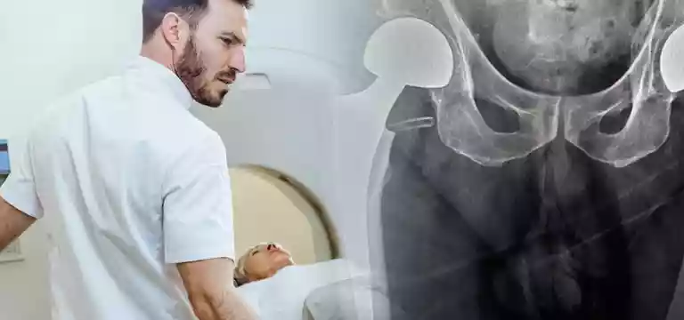
Hip pain is a common problem among the general population. In this article, we will see how magnetic resonance imaging (MRI) is the preferred diagnostic tool when comes to examining painful hip conditions, owing to its ability...
Introduction
Hip pain is a common problem among the general population. In this article, we will see how magnetic resonance imaging (MRI) is the preferred diagnostic tool when comes to examining painful hip conditions, owing to its ability to visualize multiple planes and provide excellent contrast resolution. One should know about the distinctive MRI characteristics of prevalent traumatic and pathological hip conditions.
To fully comprehend hip pathologies, it's important to have in-depth knowledge and information about the hip's anatomy.
It is important to have an overview of the key components of hip anatomy, including the bones, cartilage, labrum, joint capsule, ligaments, tendons, and bursae.
Understanding the anatomy of the hip
The hip joint, often known as a ball and socket joint, is the largest weight-bearing joint of the human body. It comprises muscles, ligaments, and tendons. The joining of the thighbone or femur with the pelvis forms this vital joint.
If the hip sustains any injury or gets inflicted by a disease, it will negatively impact its capacity for motion and weight-bearing ability.
The hip joint comprises the following components:
- Bones and joints
- Ligaments forming the joint capsule
- Muscles and tendons
- Nerves & blood vessels
Bones, joints of the hip
The hip joint connects the leg to the trunk of the body. It consists of the femur (thighbone) and the pelvis (composed of the ilium, ischium, and pubis). The femoral head forms the ball of the joint, while the acetabulum forms the socket.
The joint capsule, surrounding muscles, and ligaments help in maintaining stability. The femur has a head, neck, and trochanters, and the acetabulum is lined with a fibrocartilaginous labrum. Articular cartilage covers the weight-bearing bones, facilitating smooth and friction-free movement.
Hip joint ligaments
The hip joint is supported and stabilized by several ligaments that connect the bones including:
Iliofemoral ligament: It is a Y-shaped ligament. It connects the pelvis to the femoral head front, limiting the over-extension of the hip.
Pubofemoral ligament: A triangular ligament that joins the upper pubis to the iliofemoral ligament, attaching the pubis to the femoral head.
Ischiofemoral ligament: A group of strong fibers that originate from the ischium behind the acetabulum and blend with the joint capsule fibers.
Ligamentum teres: A small ligament extending from the tip of the femoral head to the acetabulum, containing a blood vessel that supplies the femoral head.
Acetabular labrum: It is a fibrous cartilage ring, which lines the acetabular socket. It deepens the cavity and improves the hip joint’s stability & strength.
Hip joint’s muscles and tendons
The hip joint relies on a unique combination of muscles and tendons for its function. They are:
Iliotibial band: A lengthy tendon running from the hip to the knee, serving as an attachment site for various hip muscles.
Gluteal muscles: Comprising the gluteus minimus, gluteus maximus, and gluteus medius, these muscles form the buttocks.
Adductor muscles: Found in the thighs, these muscles contribute to adduction, which involves pulling the leg back toward the midline.
Iliopsoas muscle: It is located in front of the hip joint, originating from the lower back and pelvis. It extends to the inner surface of the upper femur. It is useful for the hip’s flexion.
Rectus femoris: As the largest band of muscles situated in the front of the thigh, these muscles, also known as hip flexors, play a role in hip flexion.
Hamstring muscles: Originating at the base of the pelvis and extending down the back of the thigh, these muscles aid in hip extension by pulling it backward, as they cross the back of the hip joint.
Hip Joint’s nerves and arteries
Within the hip joint, there is a network of nerves that helps in the communication between the brain and the muscles, contributing to hip movement and transmitting sensory information such as touch, pain, and temperature.
Some of the important nerves in the hip region are femoral nerve and the sciatic nerve. Moreover, the obturator nerve provides innervation to the hip.
Along with the nerves, there are blood vessels, which are responsible for supplying the lower limbs with oxygenated blood. Among them, the femoral artery is one of the body's largest arteries. Located deep within the pelvis, it can be palpated in the front of the upper thigh.
The different anatomical components of the hip work in tandem to help in a range of movements. These movements are flexion, extension, abduction, adduction, circumduction, and rotation of the hip joint.
MRI of the hip joint
Among the masses, hip pain is seen as a widespread issue that can result from various causes. It is important to note that in diagnosing hip conditions, imaging plays a crucial role because the hip joint is very difficult to examine directly. One preferred choice and effective imaging technique is MRI imaging (MRI), which offers multiple views, excellent contrast resolution, and does not involve any harmful radiation.
MRI can help in the diagnosis of common hip conditions and identify their unique features.
Hip MRI price
Since MRI is a sophisticated diagnostic modality, it is very costly too. Compared to other image techniques, an MRI procedure tends to be more expensive. Hence, some patients may have concerns about a hip MRI price.
It is important to understand that the price of a hip MRI can vary and is dependent on numerous factors such as the healthcare facility, location of the facility, and any additional services required in a particular case. The hip MRI price may also depend on whether contrast dye is used during the scan.
So, the best you can do is to consult your healthcare provider or insurance company to get an accurate estimate of the hip MRI price and to understand any potential coverage or financial assistance options available. This will give you a clear idea and peace of mind if you are scheduled for a hip MRI.
Conclusion
We have seen that a hip MRI is a valuable diagnostic tool that provides detailed imaging of the hip joint, helping doctors and healthcare professionals to identify and evaluate various conditions and injuries related to the hip.
While the cost of a hip MRI can vary, it is important to consider the potential benefits it offers in terms of accurate diagnosis and appropriate treatment planning. Investment in an MRI can lead to improved outcomes, timely interventions, and enhanced overall patient care. So, you must immediately consult with your doctor, your insurance company, or medical facilities to obtain specific information about the cost and potential financing options available for a hip MRI. By making timely decisions, patients can take proactive steps towards better hip health and well-being.
FAQs
What is the normal cost of a hip MRI?
The cost of a hip MRI can vary based on several factors such as the location, healthcare facility, additional services needed for it, etc. It's best to consult with your healthcare provider or contact the specific imaging center for an accurate estimate.
Does insurance cover the cost of a hip MRI?
Insurance coverage for a hip MRI varies depending on the specific insurance plan you have. Some plans may fully or partially cover the cost, while others may ask for prior authorization or have certain limitations. So, contact your insurance provider to understand everything clearly.


