F-Dopa PET scan also known as 18F- dihydroxyphenylalanine PET scan/test is a diagnostic imaging modality for identification and evaluation of many pathological conditions of brain especially neurodegenerative disorders of brain.
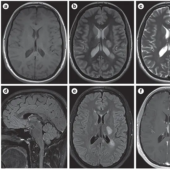
Magnetic resonance imaging (MRI) is a non-invasive and widely used medical imaging technology that leverages a strong magnetic field, radio waves, and sophisticated computing techniques to create highly detailed images of internal structures within the body. Head MRI scans, in particular, play a critical role in the diagnosis and treatment of numerous medical conditions, enabling clinicians to examine the brain, surrounding tissues, and blood vessels with great precision.
This article presents a comprehensive guide to head MRI scans, providing a detailed overview of the indications, procedure, and completion of this important diagnostic tool.
Indications for Head MRI Scans
There are several reasons why a healthcare provider might recommend a head MRI scan. The most common conditions that could require head MRI are as follows:
Headaches - Headaches are a common symptom of many medical conditions, including migraines, sinusitis, and brain tumors. An MRI scan can help identify the underlying cause of the headaches, which can guide treatment.
Traumatic Brain Injury - A head MRI scan can be used to assess the severity of a traumatic brain injury (TBI) and monitor the patient's recovery.
Seizures - Seizures are caused by abnormal electrical activity in the brain and can be caused by various medical conditions. An MRI scan can help identify the underlying cause of the seizures, which can guide treatment.
Tumors - An MRI scan can detect the presence of brain tumors and help determine their size, location, and type.
Strokes - An MRI scan can detect signs of a stroke, such as a blood clot or bleeding in the brain. Early detection is essential to minimize the damage caused by a stroke.
Multiple Sclerosis - Multiple sclerosis (MS) is a chronic disease that affects the nervous system. An MRI scan can detect the characteristic lesions in the brain and spinal cord that are associated with MS.
Preparing for a Head MRI Scan
Before the MRI scan, the patient will need to remove any metal objects from their body, such as jewelry, watches, and hearing aids. The magnetic field produced by the MRI machine can cause these objects to move or heat up, which can be dangerous.
The patient will need to change into another gown given from diagnostic centre if applicable. If the patient is claustrophobic or anxious about the scan, they may be offered a mild sedative to help them relax.
During the Head MRI Scan
Preparation : Once the patient is prepared, they will be asked to lie down on a narrow table that slides into the MRI machine. The machine is a large, tube-like structure with a magnetic field that is several times stronger than the Earth's magnetic field.
Communication : The MRI technician will communicate with the patient through an intercom system during the scan. The patient will need to remain still during the scan, as any movement can blur the images.
During Scanning : The MRI machine will produce loud thumping and tapping noises during the scan. The patient will be given earplugs or headphones to wear to block out the noise. They can also listen to music or an audiobook to help pass the time.
Time: The scan can take anywhere from 30 minutes to an hour, depending on the type of images needed. If the patient is having multiple scans, they may need to reposition themselves between scans.
Contrast Agent Injection : If the healthcare provider has ordered a contrast-enhanced MRI, the patient will be given an injection of a contrast agent before the scan. The contrast agent helps to highlight certain structures in the brain and can improve the image quality.
After the Head MRI Scan
Once the scan is complete, the patient can change back into their clothes and resume their normal activities. There are no restrictions on diet or activity after the scan, and the patient can usually drive home or return to work immediately.
The images from the scan will be sent to a radiologist, a specialist who interprets medical images. The radiologist will analyze the images and send a report to the healthcare provider who ordered the scan.
Risks and Side Effects of Head MRI Scans
Head MRI scans are generally considered safe, and there are no known risks associated with the magnetic fields and radio waves used in the scan. However, there are a few things to keep in mind:
Claustrophobia - The enclosed space inside the MRI machine can be uncomfortable or frightening for some people, especially those with claustrophobia. If the patient is anxious or claustrophobic, they can ask for a mild sedative to help them relax.
Gadolinium Contrast Agents - In rare cases, the use of contrast agents can cause an allergic reaction. Symptoms of an allergic reaction include hives, itching, and difficulty breathing. If the patient experiences any of these symptoms during or after the scan, they should notify the MRI technician or healthcare provider immediately.
Metal Implants - Patients with metal implants, such as pacemakers or cochlear implants, may not be able to have an MRI scan. The strong magnetic field can cause the metal to move or heat up, which can be dangerous.
Noise - The MRI machine produces loud thumping and tapping noises during the scan. The patient will be given earplugs or headphones to wear to block out the noise.
Anxiety - Some patients may feel anxious or claustrophobic during the scan. If the patient is feeling anxious or uncomfortable, they can ask the MRI technician for a break or a mild sedative to help them relax.
Conclusion
A head MRI scan is a non-invasive imaging test that uses powerful magnets and radio waves to produce detailed images of the brain, blood vessels, and surrounding tissues. The scan can help diagnose a range of conditions, including tumors, strokes, and traumatic brain injuries.
Before the scan, the patient will need to remove any metal objects from their body and change into a hospital gown. During the scan, the patient will need to lie still on a narrow table inside the MRI machine, which can be uncomfortable or frightening for some people. The scan can take anywhere from 30 minutes to an hour, depending on the type of images needed.
Head MRI scans are generally considered safe, although patients with metal implants may not be able to have the scan. The use of contrast agents can also cause rare allergic reactions, and some patients may experience anxiety or claustrophobia during the scan.
If you are scheduled for a head MRI scan, it is important to follow the preparation instructions given by your healthcare provider and inform them of any medical conditions or implanted devices you have.
By understanding what to expect during the scan, you can feel more confident and relaxed about the procedure. If you have any concerns or questions about the scan, you can talk to your healthcare provider, who can provide you with more information about the procedure, including the risks and benefits, and help you make an informed decision about whether the scan is right for you.


