Book Polarizing Microscopy Body Fluids Appointment Online Near me at the best price in Delhi/NCR from Ganesh Diagnostic. NABL & NABH Accredited Diagnostic centre and Pathology lab in Delhi offering a wide range of Radiology & Pathology tests. Get Free Ambulance & Free Home Sample collection. 24X7 Hour Open. Call Now at 011-47-444-444 to Book your Polarizing Microscopy Body Fluids
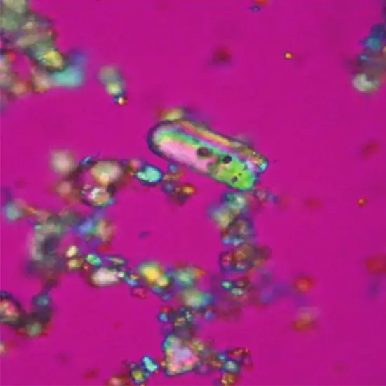
Polarized light microscopy is a contrast-enriching technique that can be operated to examine the configuration of the object. Polarized light microscopes have increased sensitivity and can be employed for quantitative and qualitative examinations. Polarized light microscopy is utilized to examine the anisotropy of a specimen's optical characteristic, which is generally induced by stress and deterioration of initially isotropic materials. The specimens could be crystalline materials (pigments, minerals, etc.) and fibers, similar to environmental particulates and natural materials.
Polarized light microscopes are traditional standard microscopes qualified with two additional polarizing components (the polarizer and the analyzer) that access sample analysis under polarized light
Pre-test Information
A last report(s), clinical description, and other components are to be proposed.
Polarising Microscopy
This possesses a foreign body in the skin, precipitating diseases, metabolic ailments, ailments of the hair, and genetic illnesses.
On polariscope analysis, distinct foreign bodies have characteristic formations. They can be established by noticing unfamiliar body granulomas (both allergic and non-allergic varieties) and most usually within multinucleated giant cells.
Cholesterol esters are doubly refractile and can be ascertained in circumstances for example tuberous xanthoma, plane xanthoma, and xanthelasma while they are passed over in eruptive xanthomas. They can also be involved in non-X-histiocytosis, juvenile xanthogranuloma, erythema elevatum diutinum, and dermatofibroma in which residue of lipids is a secondary spectacle. To define the lipids in the regions, tissue should be fixed in formalin and it should be stable.
Amyloid elements exhibit a brick red color with apple green birefringence under the polarising microscope when stained with Congo red. This unique dyeing is due to the beta-pleated sheet conformation of the polypeptide spine of amyloid fibrils. In the option of macular and lichen amyloidosis, it seems that just in the papillary upper layer while in systemic amyloidosis it also seems in the deeper layer, subcutaneous tissue, and within the cell lining of the blood vessels.
Polariscopic estimation of the hair in trichothiodystrophy presents remarkable alternate dark and white lines of hair shaft quoted a ′tiger tail′ figure which is not noticed in the light microscopic investigation.
In Fabry, ′s illness confirmations of glycosphingolipids can be predicted in the endothelial, perithelial, and smooth muscle cell border of blood vessels and urinary residues where they are marked as birefringent ′Maltese crosses′.
| Test Type | Polarizing Microscopy Body Fluids |
| Includes | Polarizing Microscopy, Body Fluids Test (Rheumatology) |
| Preparation | |
| Reporting | Within 24 hours* |
| Test Price |
₹
|

Early check ups are always better than delayed ones. Safety, precaution & care is depicted from the several health checkups. Here, we present simple & comprehensive health packages for any kind of testing to ensure the early prescribed treatment to safeguard your health.