Book Obstetric Ultrasound Appointment Online Near me at the best price in Delhi/NCR from Ganesh Diagnostic. NABL & NABH Accredited Diagnostic centre and Pathology lab in Delhi offering a wide range of Radiology & Pathology tests. Get Free Ambulance & Free Home Sample collection. 24X7 Hour Open. Call Now at 011-47-444-444 to Book your Obstetric Ultrasound at 50% Discount.
What is the Obstetric Scan Pregnancy Test?
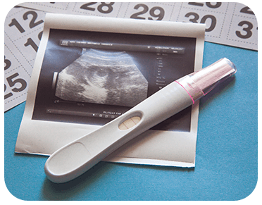
Ultrasound waves are used in obstetric ultrasound to create images of a baby (embryo or fetus) inside a pregnant woman's uterus and ovaries. There are no adverse side effects, and it is the recommended way to keep track of pregnant mothers and their unborn children.
Doctors recommend this Obstetric Scan Pregnancy Test to pregnant women for the following purposes:
The scan includes sound waves to create an image of the fetus, which is then checked for defects and anomalies. The sonographer lays the transducer on the patient's body and slides it back and forth over the target area until the appropriate images are captured.
It has no adverse effects on either the mother or the baby, making it a safe procedure to undergo.
The cost could range from INR 2000 to INR 4000 for obstetric Scan Pregnancy test in Delhi. The test should be done at a licensed diagnostic centre.
To provide hassle-free diagnostic service we also provide online service to book your appointment. At Ganesh Diagnostic, you can book Obstetric Ultrasound tests online by scheduling your appointment. Schedule now for a comprehensive health assessment. We are 24/7 Available at your service.
| Test Type | Obstetric Ultrasound |
| Includes | Obstetric Ultrasound Scan (Ultrasounds) |
| Preparation |
|
| Reporting | Within 3-4 hours* |
| Test Price |
₹ 2000
|

The 1st sign of early pregnancy seen on ultrasound scan is the presence of gestational sac. It is a spherical, fluid containing sac around the developing embryo. On ultrasound scan, an intrauterine gestational sac appears as two concentric echogenic rings that are separated by a hyperechoic space. This finding is known as Double decidual sac sign and indicates a normal intrauterine pregnancy.
Gestational sac can be seen as early as:
6 weeks of gestation by Transabdominal ultrasound/obstetric ultrasound scan.
There is no difference between sonography and an ultrasound scan and both the terms can be used interchangeably. However, the term sonogram refers to image produced by an ultrasound scan or sonography test.
Normally 2-3 ultrasound scans are performed during pregnancy. However, if required multiple scans can be done. According to ACOG (American college of obstetricians and gynaecologist), ultrasounds are safe during pregnancy and there is no evidence demonstrating that ultrasonography pose any kind of risk to the growing foetus.
In the first trimester of pregnancy, following ultrasound scans can be done:
Level 2 ultrasound is performed in the second trimester during 18-22 weeks of pregnancy. It is also known as Anomaly scan or TIFFA scan. It is a mandatory scan recommended for every pregnant woman.
Level 2 ultrasound or TIFFA scan is used to detect congenital malformations, growth abnormalities in the fetus as well as identify placental pathologies.
In the third trimester of pregnancy, following ultrasound scans can be done:
Obstetric Color doppler also known as color doppler ultrasound in pregnancy is a diagnostic tool that is used in pregnancy to check the patency, direction and speed of blood flow in maternal as well as fetal blood vessels. It is commonly done in third trimester of pregnancy. However, in high-risk pregnancies it may be done at an earlier stage. It also provides guidance in deciding time of delivery in such cases.
Color Doppler ultrasound is a non-invasive, painless and fast modality of imaging which is safe in pregnancy as it doesn’t utilize ionizing radiations or contrast agents rather it utilizes sound waves for depicting any underlying pathology of maternal or fetal blood flow.
Some of the indications/uses of color doppler ultrasound in pregnancy includes:
It’s not very difficult to read or understand ultrasound reports of pregnancy. However, the only pre-requisite is your basic understanding of some medical terms/terminology.
You will receive the reports with description of your ultrasound findings along with attached picture/sonogram. You may read and understand the printed description of your ultrasound findings but you may not completely understand the sonogram attached with it.
Usually, the sonogram is provided for your obstetrician reference purpose. So that she may look and analyse the co-relation between the description provided and the sonogram attached with it. This helps your obstetrician in understanding and developing proper management plan according to your current scenario.
Sometimes urine pregnancy tests are positive but when you go for an obstetric ultrasound scan it turns out to be negative for pregnancy. This is a common finding encountered by many women. Various reasons or causes of this finding includes the following-
Ultrasound is a painless, non-invasive and rapid technique to confirm pregnancy, evaluate gestational age and EDD (expected date of delivery). The presence of gestational sac on USG scans is considered as the 1st definitive sign of pregnancy and the presence of yolk sac within the gestational sac is considered as the 1st reliable sign of intrauterine pregnancy.
The best time to calculate gestational age of baby is the 1st trimester of pregnancy (9-12 weeks) by an ultrasound scan (using the parameter CRL). Gestational age is calculated using CRL (Crown-rump length) in the 1st trimester of pregnancy as following-
However, if pregnant women present for the first time in 2nd trimester of pregnancy, then the parameters used in ultrasonography to calculate gestational age of foetus is femur length (FL), Head circumference (HC), Biparietal diameter (BPD) or Abdominal circumference (AC).
And if pregnant women present for the 1st time in 3rd trimester of pregnancy, then parameters used for the estimation of gestational age includes femoral epiphyseal ossification centers, proximal tibial ossification centers or proximal humeral ossification centers.
Obstetric Ultrasound usually takes around 15-30 minutes. There are no known side effects of Obstetric Ultrasound and this procedure is safe.
Patients can visit the Ganesh diagnostic website for Obstetric Ultrasound reports or can get through a registered mobile number.
Patients can type Obstetric Ultrasound centres near me in Delhi NCR in Google search for the nearest centres available.
Early check ups are always better than delayed ones. Safety, precaution & care is depicted from the several health checkups. Here, we present simple & comprehensive health packages for any kind of testing to ensure the early prescribed treatment to safeguard your health.