Book FISH MET Amplification Appointment Online Near me at the best price in Delhi/NCR from Ganesh Diagnostic. NABL & NABH Accredited Diagnostic centre and Pathology lab in Delhi offering a wide range of Radiology & Pathology tests. Get Free Ambulance & Free Home Sample collection. 24X7 Hour Open. Call Now at 011-47-444-444 to Book your FISH MET Amplification at 50% Discount.
The receptor kinase MET protein, like EGFR, promotes tyrosine-kinase activity. Signaling pathways implicated in cell growth and survival are turned on when tyrosine kinase is promoted. As a result, non-small-cell lung cancer patients with MET protein overexpression, MET proto-oncogene amplification, and MET exon 14 skipping mutation have tumor development and progression with a poor prognosis (NSCLC).
When EGFR-activating modifications are present in people with NSCLC, MET amplification is connected to attained resistance to EGFR tyrosine kinase inhibitor (TKI) therapy. In NSCLC cases with high-level MET amplification or MET exon 14 omitting modification, crizotinib (see NCCN recommendations) or another MET-targeted treatment may be taken into thinking.
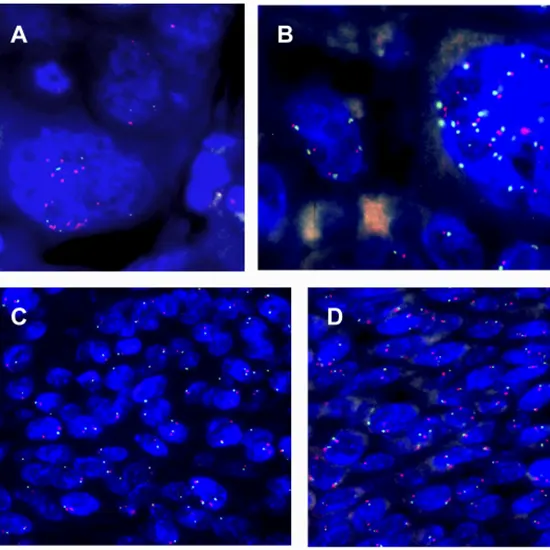
To assess copy number gain in interphase cells from the target lesion, the MET (7q31) FISH dual-color probe set is hybridized with a control probe (D7Z1).
Coexisting MET mutations and other oncogene mutations are possible (eg, KRAS, EGFR, BRAF, ALK). MET overexpression and/or amplification are linked to a more virulent illness, just as mutations in these other oncogenes.
An average of two MET signals and two control probe (D7Z1) signals should be present in healthy tissues. Copy number gain is indicated by the over-representation of MET in comparison to the control.
MET copy quantity per cell, MET/D7Z1 ratio, and percentage (%) of positive cells make up the scoring system.
Fresh tumor tissue as well as formalin-fixed, paraffin-embedded tissue can be evaluated.
The IHC assay determines the degree of gene expression, whereas the FISH assay determines the number of copies of the gene (i.e., MET amplification) (ie, MET overexpression). Even though FISH and IHC results typically correspond, NCCN recommendations only suggest FISH testing for MET amplification.
Take tissue from the tumor.
Tissue from the tumor is paraffin entrenched and fixed with formalin (10 percent neutral buffered formalin). Ferry tissue block or four successively cut, 5-micron-thick, unstained pieces put on glass slides with a positive charge. (4 slides lowest) Keep slides and/or paraffin blocks away from extreme heat.
Storage/Transport Temperature
Ambient temperature. Also acceptable is chilled.
Specimens treated or repaired with heavy metal fixatives or alternative fixatives (alcohol, Prefer) (B-4 or B-5). tissue free of tumors. skeletal specimens.
| Test Type | FISH MET Amplification |
| Includes | FISH MET Amplification (Pathology Test) |
| Preparation | |
| Reporting | Within 24 hours* |
| Test Price |
₹
|

Early check ups are always better than delayed ones. Safety, precaution & care is depicted from the several health checkups. Here, we present simple & comprehensive health packages for any kind of testing to ensure the early prescribed treatment to safeguard your health.