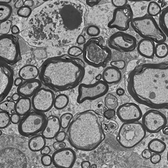
Book Electron Microscopy Appointment Online Near me at the best price in Delhi/NCR from Ganesh Diagnostic. NABL & NABH Accredited Diagnostic centre and Pathology lab in Delhi offering a wide range of Radiology & Pathology tests. Get Free Ambulance & Free Home Sample collection. 24X7 Hour Open. Call Now at 011-47-444-444 to Book your Electron Microscopy at 50% Discount.
Electron microscopy is used to obtain ultrahigh-resolution images of individual atoms of materials of cells. These resulting microstructure and mesostructure images are usually used to investigate the sample properties and behavior.
These electron microscopes produce electron micrographs using technical digital cameras and frames to capture the images.
The science of microbiology exhibits its development to the electron microscope. The study of microorganisms like bacteria, virus, and other pathogens by electron microscopes have made the treatment of diseases very effective.
Scanning electron microscopy (SEM) uses a relatively low-power electron beam for imaging and interaction with the sample.
Transmission electron microscopy (TEM) uses a high-energy beam of electrons to transmit electrons through a sample to create a 2D image at the highest possible resolution.
An electron microscope is an instrument that uses a beam of electrons to magnify a specimen. It is mainly used to observe the internal structure of cells and the ultrastructure of surfaces.
It is used in conjunction with a variety of ancillary techniques (e.g. thin sectioning, immuno-labeling, negative staining) to answer specific questions. EM images provide key information on the structural basis of cell function and of cell disease.
It is used in materials science, biomedical research, quality control, and failure analysis. The use of electrons as the imaging radiation source allows for greater spatial resolution (on the tens of picometers scale) as compared to the resolution achieved using photons in optical microscopy. In addition to surface topography, information about crystalline structure, chemical composition, and electrical properties are obtainable through electron microscopy.
Electron microscopes always occupy signals generated from the interaction of an electron beam with the sample to obtain information about structure, morphology, and composition.
The difference between a regular microscope and an electron microscope is that it uses a beam of electrons to produce images. A regular microscope uses light to magnify a specimen.
| Test Type | Electron Microscopy |
| Includes | Election Microscopy (Lab Test) |
| Preparation | |
| Reporting | Within 24 hours* |
| Test Price |
₹ 3500
|

Early check ups are always better than delayed ones. Safety, precaution & care is depicted from the several health checkups. Here, we present simple & comprehensive health packages for any kind of testing to ensure the early prescribed treatment to safeguard your health.