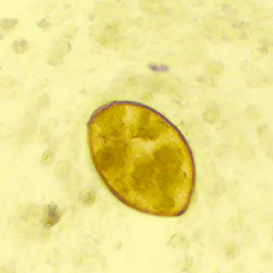
Book Direct Microscopy For Paragonimus Westermani Appointment Online Near me at the best price in Delhi/NCR from Ganesh Diagnostic. NABL & NABH Accredited Diagnostic centre and Pathology lab in Delhi offering a wide range of Radiology & Pathology tests. Get Free Ambulance & Free Home Sample collection. 24X7 Hour Open. Call Now at 011-47-444-444 to Book your Direct Microscopy For Paragonimus Westermani at 50% Discount.
Following centrifugation, specimens of Paragonimus westermani are inspected under a microscope for eggs using the Formalin/Ethyl-Acetate Concentration Procedure.
A parasitic worm infestation is known as paragonimiasis. Eating crab or crayfish that are not fully cooked is the cause.
Paragonimiasis may cause pneumonia or symptoms like stomach flu. An infection could linger for years.
Paragonimiasis develops when a flatworm infection is present. That parasitic worm is also called a fluke or lung fluke because it frequently attacks the lungs. Consuming raw crab or crayfish that include young chances often leads to disease.
After being ingested by a person, the worms develop and grow inside the body. For several months, the worms spread throughout the stomach and intestines (abdomen). They pass through the diaphragm muscle to get to the lungs. In the lungs, the worms can lay eggs and live for many years, causing chronic (long-term) paragonimiasis.
During the earliest stages of infection, paragonimiasis has no symptoms. Many persons who have paragonimiasis show no signs at all. When paragonimiasis symptoms appear, they result from the worms' shifting activity and position within the body.
Within the first month or two following infection, paragonimiasis worms spread throughout the belly, perhaps leading to symptoms like:
After that, worms go from the stomach into the chest. There, they may induce respiratory issues like:
The use of electron microscopy allows for the creation of ultrahigh-resolution images of individual material or cell atoms. Oftures of the microstructure and mesostructure produced as a consequence are typically used to examine the characteristics and behaviour of the sample.
Modern microscopes create electron micrographs by capturing images with sophisticated digital cameras and frames.
The progress of microbiology is demonstrated via the electron microscope. By using electron microscopes to investigate microorganisms like bacteria, viruses, and other pathogens, diseases can be treated quite successfully.
There are two primary forms of electron microscopy:
A relatively low-power electron beam is used in scanning electron microscopy (SEM) for imaging and interacting with the material.
A high-energy electron beam is used in transmission electron microscopy (TEM) to transmit electrons through a sample and produce the highest-resolution 2D image possible.
| Test Type | Direct Microscopy For Paragonimus Westermani |
| Includes | Direct Microscopy For Paragonimus Westermani (Pathology Test) |
| Preparation | |
| Reporting | Within 24 hours* |
| Test Price |
₹ 110
|

Early check ups are always better than delayed ones. Safety, precaution & care is depicted from the several health checkups. Here, we present simple & comprehensive health packages for any kind of testing to ensure the early prescribed treatment to safeguard your health.