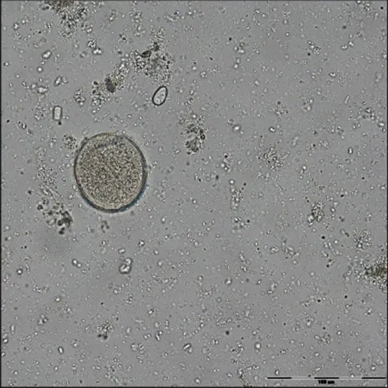
Protozoan parasitism encompasses a wide range of diseases. Balanthidium coli and balantidiosis, the subjects of this review, are rarely included in infectious protozoal diseases.
Protozoan parasitism encompasses a wide range of diseases. Balanthidium coli and balantidiosis, the subjects of this review, are rarely included in infectious protozoal diseases.
Balantidium is the only ciliated protozoan known to infect humans and is the largest protozoan to infect humans and non-human primates. Balantidiosis is a zoonotic disease that is transmitted to humans via the fecal-oral route from the usual host pigs and is asymptomatic.
Water is the carrier in most cases of balantidiosis. Human-to-human transmission is also possible. The human Valandidium habitat is the cecum and large intestine. Humans, like pigs, can remain asymptomatic or develop dysentery similar to Entamoeba histolytica.
Death is a rare consequence of balantidiosis, but in developing countries with malnourished and hyperparasitized populations, it can mean the difference between a healthy life and chronic wasting.
Contaminated with pig and human excrement. B. coli can be an opportunistic parasite in immunocompromised hosts living in urban environments where pigs are not the source of infection.
Balantidium is an often neglected pathogen. Research on barantidium is sparse. Zaman said that 30 years ago he published a comprehensive review of Balantidium, but recently the organism was considered an emerging protozoan pathogen and was reviewed by Garcia.
Morphology
Vegetative ciliates are 30–150 μm long and 25–120 μm wide. The spherical or slightly ovoid cysts are 40–60 μm in diameter. However, they vary in size, some up to 200 μm in length. The mouth (oral apparatus) is at the tapered anterior end and the cell division (anus) is at the rounded posterior end.
The cytoplasm has a sausage-shaped macronucleus and a rounded micronucleus. Asexual division occurs as in most ciliates. A lateral groove forms that divides the mother cell into two asymmetric daughter cells, the anterior (proter) and posterior (opisthe) cells. Porter keeps oral appliances and opisthe develops new appliances.
Areas of anterior mouth formation appear in opistes, leading to the formation of new oral apparatus, but old oral apparatus may also undergo reorganization. Swimming organisms exhibit rotational movement by somatic cilia, facilitating movement through colonic contents.
Sexual reproduction as conjugation of balantidium has been reported, but information on the details of the nuclear event is lacking. Two sequential breaks (meiosis) occur, preceded by the formation of zygotes by equal or unequal divisions. The two conjugates attach to the oral apparatus and exchange meiotic micronuclear products.
Physiology
Few studies have examined the energy metabolism of these organisms. Balantidia can survive in both anaerobic and aerobic conditions. Carbohydrates are the main source of energy for ciliates growing in vivo.
In an E. coli study combining ultrastructural and cytochemical studies, peroxisomes were identified in ciliates. These vesicles contain peroxidase, an enzyme that protects against the destructive effects of highly oxidizing compounds such as hydrogen peroxide. A comparison of ciliate cytoplasm from asymptomatic and acute barantidiosis pigs was made.
Peroxisomes were more numerous but smaller (0.6–0.8 μm) in barantidia of asymptomatic pigs than in acutely ill pigs (>0.8 μm). Similarly, the nucleic acid content (particularly RNA but also DNA) differs between symptomatic and asymptomatic pigs, with the former having a higher content.
This difference may depend on the degree of ciliate metabolic activity and, in the case of RNA, may indicate increased protein synthesis. Ciliates with higher nucleic acid content produced more CT Chest robust cultures, at least during the early stages of in vitro growth. The enzyme glucose-6-phosphatase was present in small vesicles attached to the endoplasmic reticulum or in the membrane itself.
Alkaline phosphatase is found in the ciliate cortex, nuclear membrane, ciliary membrane, kinetosome, and vesicles of the endoplasmic reticulum.
Phosphatase enzymes play an important role in providing glucose as an energy source.
Balantidia produces no known toxins, but its ability to induce colon wall ulcers is attributed to hyaluronidase, an enzyme that digests hyaluronic acid, a component of the 'glue' that holds mucosal epithelial cells together. I'm here.
Dissolution of group C streptococcal capsules containing hyaluronic acid and degradation of potassium hyaluronate by viable B. E. coli was employed as evidence for hyaluronidase activity. Entamoeba histolytica is a classic protozoan dysentery pathogen that attacks the mucosal surface of the colon and has long been claimed to possess hyaluronidase activity. However, attempts to demonstrate its existence using E. histolytica extracts did not support this claim.
Laboratory diagnosis
Due to its large size and helical movement, it is easily recognizable by wet mounting even at low magnification (100x). This is the case for freshly collected diarrheal stool specimens, which likely contain actively floating vegetative ciliates as well as bronchoalveolar lavage fluid.
Fecal samples for testing should be collected over several days, as shedding of parasites can be unstable. In formed stools, the cystic stage is more common. To check for cysts, a portion of the stool that has formed is minced with phosphate-buffered saline or a fixative (10% phosphate-buffered formalin or polyvinyl alcohol) and coarsely filtered through gauze or a sieve to remove large pieces. Remove.
The resulting fluid can be examined microscopically for formed stool cysts or diarrheal trophozoites. Phase-contrast microscopy is useful for viewing the internal structure of unstained live or fixed ciliates. The same applies to permanent dyes such as hematoxylin-eosin.Heavily stained cysts can be mistaken for helminth eggs, leading to misdiagnosis.
If a colonic biopsy was performed, hematoxylin-eosin staining of the sections will follow to help assess the extent of wall damage. Sedimentation Stool Culture and flotation methods are methods for concentrating parasites from faecal samples and making them easier to find.
Because barantidiosis is a rare disease in developed countries, most technicians do not typically look for balanthidium trophozoites or cysts when examining stool specimens.
Therefore, it is particularly important to consider the possibility of balantidiosis for patients from endemic areas and for travelers returning from such areas. He may have high numbers of Balantidia in Stool Routine stool samples. Feces of 1,230 organisms g-1 have been reported from chimpanzee faeces in Japan. A Danish study of pigs on research farms found an average of 865 cysts g-1 in the faeces of pigs aged 28 to 52 weeks or older.
Lung infection
Diagnosis of B. coli pulmonary infection can be problematic due to possible confusion between ciliated epithelial cells (CECs) and vegetative balantidia. Bronchoalveolar lavage fluids containing balantidium have been reported (3,58,70), but may also contain motile CECs from the trachea, which could be mistaken for wet mount ciliates.
CECs have relatively few cilia on their surface compared to the uniform ciliated surface of Balantidia, and in the case of columnar epithelial cells, cilia may be clumped together. CECs are smaller (<30 μm) than Valandizia (150–200 μm), oblong rather than ovoid, and swim aimlessly in circles, in contrast to the prominent spiral movements of ciliates. tend to.
Using phase-contrast microscopy helps visualize valandium features such as the oral apparatus, smooth body cilia, Ultrasound and macronuclei. Abnormal cilia were seen in wet mounts while searching the infant's nasopharynx for respiratory syncytial virus and were initially thought to be a parasite, possibly E. coli.
Subsequent examination after staining revealed a distribution of cilia along one edge of the cells, confirming that the cells were ciliated membranes, degenerate fragments of epithelial cells. CEC is sometimes confused with other motile pathogens. In this case, the flagellated stage of the amoeba Naegleria fowleri in the CSF sample.
Prevention of infection
The best way to protect mankind from valandidiosis is to provide clean, unpolluted water for drinking and other uses. Chlorine is not effective against barantidium cysts at concentrations typically used to ensure water safety.
Pigs should not be allowed to walk in or around tributaries or rivers that feed into reservoirs used for municipal water supply. May be contaminated with valandium cysts. Pigs should not have access to land where crops are grown.
Judging from the incidence of balanthidiasis in immunocompromised individuals living in urban settings, there are other sources of infection besides pig-to-human transmission. Breeding balantidium-free pigs is an unrealistic goal. Piglets are infected through their mothers or, failing that, by cannibalism.


