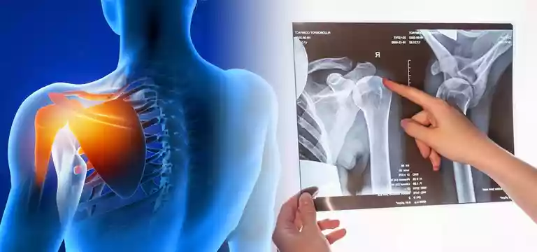
Magnetic resonance imaging (MRI) is a non-invasive, painless, and safe imaging modality, used to diagnose a wide array of diseases apart from monitoring the recovery process from surgery.
Introduction
Magnetic resonance imaging (MRI) is a non-invasive, painless, and safe imaging modality, used to diagnose a wide array of diseases apart from monitoring the recovery process from surgery. The best part is, MRI doesn’t use radiation and is devoid of any recovery time.
So, just like other body organs, an MRI is also a valuable diagnostic tool to detect problems in the shoulder.
Understanding an MRI procedure
An MRI uses magnetic fields, radio waves, and a computer to obtain detailed images of internal body parts in slices known as cross-sections. It is touted as a gold standard test for problems of the soft tissues. The MRI images showcase only a few super-thin tissue layer at a time. So, it immensely helps physicians to detect problems.
A doctor will order an MRI to assess various conditions implicating the shoulder and other joints, to get a clear visualization of soft tissues, bones, ligaments, etc. It can also help in pinpointing other disorders like:
- Degenerative joint disorders such as arthritis
- Shoulder impingement, involving pressure on tendons or nerves
- Rotator cuff injuries
- Torn ligaments
- Sports-related injuries
- Repetitive strain injuries causing pain and damage
- Bone infections
- Persistent shoulder pain unresponsive to treatment
- Limited shoulder mobility
- Monitoring shoulder healing post-surgery
- Identification of tumors
With the help of an MRI, doctors can derive crucial information to devise appropriate treatment plans and interventions for various shoulder conditions.
Know what an MRI Contrast is?
In some cases of MRI, a contrast has to be administered including a shoulder scan. Just before the start of the process, the contrast dye is injected with an intravenous needle or IV. The purpose of this contrast agent is to collect around tissues and cells so that it is easier to see parts of the shoulder. In short, the contrast agent helps in generating more clear images.
So, a doctor might do a shoulder MRI with contrast to obtain better images. There is less chance of the contrast agent causing allergic reactions compared to other types. However, one might have a cool sensation once it’s injected into the vein.
How is an MRI conducted?
During an MRI scan, the patient is asked to lie down on an examination table that slides into a long, narrow tube-like MRI machine with open ends. The MRI machine is wrapped around with a large magnet. The whole machine is a large magnet as the process is based on magnetic fields and radio waves to click images of the internal organs of the body.
After that, a trained technician will take over and operate the MRI machine from an adjacent room. The technician will closely monitor the process while capturing detailed images of the patient’s body. There is constant communication between the patient and the assigned technician with the help of a speaker attached to the machine. The patient has to follow any instructions given by the technician so that images are clear and helpful for investigation.
The MRI process is usually painless, but some people may experience feelings of nervousness or claustrophobia due to the confined space. If you have any issues and find it difficult to remain still throughout the process, talk to your doctor beforehand. The doctor may suggest a sedative so that the patient can relax and ease the anxiety. However, one should remember that only the doctor should prescribe any such medication, and the technicians are not eligible to suggest any relaxant to the patient. It is crucial to remain as still as possible during the MRI to obtain accurate images.
Getting ready for a Shoulder MRI
Moreover, there shouldn’t be any metallic objects in the body during the scan. One should inform the doctor well in advance about any metal implants in the body. Metallic objects are likely to be attracted by the strong magnetic field within the MRI machine. It will jeopardize the safety of the patient.
All these need to be discussed with the doctor beforehand. The metallic implants could be:
- Brain aneurysm clips
- Artificial heart valves
- Cochlear implants
- Pacemakers
- Artery stents
- Recent artificial joints
- Vagal nerve stimulators, etc.
Once a patient is declared ready after clearing all the above criteria, he or she will get instructions on how to get ready for the test from the imaging lab. Some patients may be asked to refrain from eating and drinking for up to four to six hours before the scan. The patient will be asked to wear comfortable clothing and remove all jewelry, hearing aids, hairpins, removable mouth appliances, etc., and keep all belongings outside the MRI machine room.
After the MRI shoulder
Once the MRI scan is over, a radiologist will carefully examine the captured images and create a report detailing any irregularities or abnormalities observed from the images. The patient will have to go for a follow-up appointment with the doctor to review the report and discuss the findings.
The radiologist will provide a description of any potential issues or abnormalities detected in the images. One must know that MRI provides cross-sectional slices of the shoulder, and hence the radiologist's interpretation may include terms like "possible tear" or "probable tear" to indicate their level of certainty.
A normal shoulder MRI result will indicate that no visible problems or abnormalities were detected during the scan. On the other hand, abnormal results will mention the presence of issues such as tears, arthritis, cysts, or other shoulder-related issues. The doctor will closely read and explain the meaning of the results and guide the patient through the appropriate next steps for treatment or further evaluation.
MRI of shoulder price
Since MRI is a highly developed and sophisticated diagnostic modality, it is a little expensive. So, many patients do have a concern about the cost. If you are looking for an MRI of the shoulder price, you must first talk to your doctor and the healthcare facility where you intend to get the test done. They will provide you with the exact price of the shoulder MRI. Remember, the MRI of shoulder price is dependent on a lot of factors including the location of the facility, the standard of the facility, the use of contrast, and the insurance coverage a patient has. One should also talk to the insurance company for the coverage policies. Even some facilities have discounted rates for patients who do not have insurance coverage. So, check with them for the final price of the shoulder MRI.
Conclusion
We now know that a shoulder MRI is an invaluable diagnostic imaging modality that provides accurate and detailed imaging of the shoulder joint, assisting doctors in precisely assessing and diagnosing various shoulder conditions. It helps in the viewing of soft tissues, bones, and abnormalities, helping doctors detect any issues like tears, arthritis, impingement, and other shoulder injuries.
Since it is a non-invasive technique MRI is a safe option for patients, and the images obtained can be thoroughly scrutinized by radiologists to provide valuable insights into the underlying causes of shoulder pain or dysfunction.
With the information obtained from a shoulder MRI, doctors can formulate appropriate treatment plans and provide patients with better shoulder health and improved quality of life.


