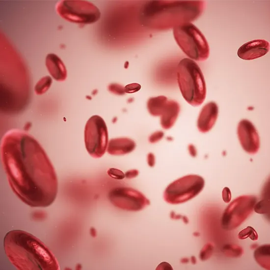
Anemia—a condition in which haemoglobin (Hb) concentration and/or red blood cell (RBC) numbers are lower than normal and insufficient to meet an individual’s physiological needs—affects roughly one-third of...
What is anemia :
Anemia- a condition in which haemoglobin (Hb) concentration and/or red blood cell (RBC) numbers are lower than normal and insufficient to meet an individual’s physiological needs- affects roughly one-third of the world’s population. Anemia is associated with increased morbidity and mortality in women and children, poor birth outcomes, decreased work productivity in adults, and impaired cognitive and behavioural development in children. Preschool children (PSC) and women of reproductive age (WRA) are particularly affected.
For anaemia to be correctly diagnosed and its detrimental effects to be avoided, suitable Hb thresholds must be established. The development of suitable interventions that address the context-specific causes of anaemia and the evaluation of the efficacy of anaemia control programmes depend on an understanding of the varied and complex aetiology of anemia, which is equally essential. To that end, the main goals of this paper are to define anaemia and categorise it, to describe the biological mechanisms by which anaemia develops, to review the various conditions and factors that contribute to anaemia development, with a focus on those that are most common in low- and middle-income countries (LMICs), and to determine the areas that require further study.
Defining anemia
Alternately, anaemia is described as a condition in which the quantity of RBCs (and consequently their capacity to carry oxygen) is insufficient to satisfy physiologic requirements.
Understanding how Hb naturally differs by age, sex, pregnancy status, genetic and environmental variables, and possibly ethnicity, is necessary to define an abnormally low Hb concentration. Hb fluctuates with age, peaking during the first few months of existence. Normal Hb concentrations are at their highest peak during life—between 17 and 21 g/L—in new-borns. The Hb concentration then drops during the first two to three months of life before rising again in childhood, then levelling off throughout maturity, before falling again as people get older. Because menstruation affects iron stores and causes anemia, sex variations in Hb concentrations start in puberty and last throughout the reproductive years. Due to the expansion of blood volume and subsequent dilution effect, Hb concentration normally decreases during pregnancy.
The sex, age, and pregnancy-specific WHO Hb cutoffs for anaemia are commonly used throughout the world. These cutoffs were initially developed in 1968 by a nutritional anaemia research group at the WHO using statistical cutoffs as opposed to thresholds connected to significant health outcomes. Since then, the 25 Hb cutoffs have been slightly altered to account for new child age groups, adjustments for children in the 5–11 age range based on non-iron-deficient children from the United States, and the development of "mild," "moderate," and "severe" anaemia categories.
Results from participants in the Second National Health and Nutrition Examination Survey (NHANES II) who were not iron deficient also validated the cutoffs.
Global magnitude of anemia
In 2010, it was predicted that anaemia affected about 32.9% of the world's population. Children under the age of five (42 percent had anaemia in 2016), especially infants and young children under two years old, WRA (39 percent had anaemia in 2016), and pregnant women (46 percent had anaemia in 2016) are the demographic groups most at risk for anaemia. In almost all age groups and geographic areas, women continually had a higher anaemia risk than men did. The elderly are another group that is at risk, as the prevalence of anaemia increases with age in people over 50,35 though the available information is sparse.
Anemia reduction has generally progressed slowly and unevenly. Anemia is estimated to have decreased approximately seven percentage points between 1990 and 2016, from 40% to 33%, for all age groups and both sexes. By 2025, the WHO Global Nutrition Target for anaemia seeks to cut anaemia in WRA by 50%. To reach this goal, a reduction of 1.8–2.4 percentage points per year would be needed, based on a worldwide prevalence of anaemia among WRA (non-pregnant and pregnant, respectively) of 29–38% as of 2011.
Additionally, geographical differences in anaemia incidence exist. In 2010, the regions with the highest anaemia prevalence across all age categories and both sexes were Sub-Saharan Africa, South Asia, the Caribbean, and Oceania.2 In the majority of WHO member states, anaemia among WRA and children under the age of 5 is a moderate-to-severe public health problem (20% or higher as defined by WHO).
Pathophysiology of anemia
Anemia has detrimental effects on both health and development results. These effects include decreased oxygen delivery to tissues, which may have an impact on multiple organ systems, as well as complications from the anaemia's complex underlying causes. For instance, decreased iron availability in iron deficiency anaemia (IDA) has well-documented detrimental impacts on brain development and functioning even before anaemia develops.
Etiology of anemia
- Anemia arises biologically from an imbalance between erythrocyte production and loss, which can be brought on by inadequate or inefficient erythropoiesis (caused, for example, by nutritional deficiencies, inflammation, or genetic Hb disorders) and/or excessive erythrocyte loss. (Due to hemolysis, blood loss, or both).
- Additionally, because anaemia can have numerous causes, even in the same person, the haematological symptoms of one cause may be concealed by those of another. For instance, macrocytic anaemia is a sign of anaemia brought on by vitamin B12 or folate deficiency. The consequences of a B12 or folate deficiency may completely be hidden by concurrent ID, which results in microcytosis. Although there are indices used in clinical practise to differentiate the causes of anaemia based on RBC parameters (e.g., IDA versus -thalassemia, which both produce hypochromia and microcytosis), their accuracy in doing so varies.
- Poor socioeconomic standing is associated with a higher chance of anaemia in women and children, for instance, and is a significant determinant of health and nutrition.
- Malaria is frequently cited as a major cause of severe anemia, especially in African children, even though studies on its aetiology are scarce. Malaria, poor sanitation, underweight, and inflammation were the most reliable predictors of severe anaemia in population-based surveys of PSC in the BRINDA project (only in African countries); stunting, vitamin A deficiency (VAD), and rural location were also significant predictors in high/very high infection countries. Malaria, bacteraemia, hookworm infection, HIV infection, a lack of glucose-6-phosphate dehydrogenase (G6PD), and deficiencies in vitamin A (VA) and B12 were all found to be linked with severe anaemia in a study of Malawian PSC.
Nutritional anaemias:
- Iro
- Vitamin B12
- Vitamin A
- Riboflavin
When hematopoietic nutrient concentrations—those involved in RBC production or maintenance—are inadequate to meet those needs, nutritional anaemias occur.13 Inadequate dietary intake, increased nutrient losses (such as haemorrhage from parasites, childbirth, or heavy menstrual losses), impaired absorption (such as a lack of intrinsic factor to aid vitamin B12 absorption, a high phytate intake, or Helicobacter pylori infection that impairs iron absorption), or altered nutrient metabolism are all causes of nutrient deficiency. (e.g., VA or riboflavin deficiency affecting mobilisation of iron stores.
Iron deficiency anaemia:
- When dietary iron intake is insufficient over time, notably during times of high iron demand (e.g., during periods of rapid growth and development, such as infancy and pregnancy), or when iron losses outweigh iron intake, ID develops.
typically evolves in three stages: storage iron depletion, iron-deficient erythropoiesis, and IDA (defined as concomitant ID plus anemia). The WHO recommends assessing iron status using serum ferritin or soluble transferrin receptor (sTfR). Serum ferritin, a measure of body storage iron and a sensitive measure of ID, is elevated by the acute phase response; sTfR levels when high indicate tissue ID, but sTfR may also be affected by inflammation and other causes of erythropoiesis. Because of the effect of inflammation on many biomarkers of iron status, acute phase proteins (e.g., C-reactive protein (CRP) and alpha-1-acid glycoprotein (AGP)) should be assessed when possible.
The WHO estimated the "proportion of all anaemia amenable to iron" as 50% of anaemia among non-pregnant and pregnant women, and 42% of anaemia in children based on the shift in Hb concentration from iron supplementation trials.9 Though the percentage of anaemic children and women with ID varied by the burden of infectious diseases, recent analyses from the BRINDA project showed that along with age and malaria, ID was one of the variables most consistently associated with anaemia.
Vitamin B12
- Riboflavin (B2), pyridoxine (B6), cobalamin (B12), and folate are some of the B vitamins that are involved in the production of haemoglobin or the breakdown of iron. Anemia has been linked to nutritional deficiency in these substances.
Macrocytic anaemia can result from a lack of folate or vitamin B12 (cobalamin). Lack of these nutrients can cause megaloblastic changes, such as hyper-segmented neutrophils on a blood smear, which impact DNA synthesis and cell division in the bone marrow.70 Erythrocyte life span can be shortened because of folate insufficiency. Low dietary consumption of the nutrient is the main cause of vitamin B12 deficiency in LMIC.
Its bioavailable forms are only present in animal-source foods, but vitamin B12 deficiency can also be caused by malabsorption, especially in elderly people with common gastric atrophy, in people with pernicious anemia, an autoimmune condition where autoantibodies are formed against an intrinsic factor necessary for B12 absorption, and in people with bacterial and parasitic infections.
Riboflavin
- A riboflavin deficiency in animals can reduce iron mobilisation from stores, decrease iron absorption, increase iron losses, and impede globin production. Riboflavin plays a crucial function in iron metabolism as a cofactor in redox reactions.55 In both high-income and LMICs, riboflavin deficiency has been documented in pregnant and lactating women, infants, school-age children, adolescent girls, and the elderly, particularly where consumption of milk/dairy products and meat (the primary riboflavin sources) is low.
Uncertainty exists regarding whether riboflavin insufficiency is a major cause of anaemia in people. Some studies, but not all, have found that riboflavin supplements given along with iron supplements have a higher impact on Hb concentration in children and pregnant women than iron supplements alone.
Vitamin A
- VAD and anaemia have been observed to coexist in the same communities for decades, and substantial correlations between VA status biomarkers and Hb have been reported in numerous nations and populations, including children of preschool and school age, adolescents, and adults.
Multiple mechanisms, including the function of retinoids in erythropoiesis, the significance of VA for immune function, and VA's well-known function in iron metabolism, are believed to contribute to anaemia in VAD. Anemia caused by VAD is distinguished from anaemia caused by IDA by an increase in iron stores in the liver and spleen as well as a rise in serum ferritin levels.
Other conditions related to anaemias
- Undernutrition
- Overweight /Obesity
Some research, but not all, have linked anaemia with stunting, wasting, and underweight. Poor maternal nutrition, inadequate home and community environments, inadequate complementary feeding practises leading to poor micronutrient and animal-source food intake, contaminated water and poor sanitation, suboptimal breastfeeding practices, clinical and subclinical infections, and other similar factors (though not constituting a causal relationship) are associated with these manifestations of poor nutritional status and anaemia.
Hepcidin, a peptide hormone primarily produced by the liver and in charge of maintaining iron homeostasis, is believed to be the main factor connecting these conditions. Hepcidin levels are elevated in an inflammatory environment. Reduced iron absorption is caused by a rise in hepcidin levels brought on by the chronic subclinical inflammation that is common in overweight and obese people. However, Hb levels typically fall within the usual range.
Summary
Anemia is still a major global health issue that needs to be properly addressed, especially in LMICs where development has been uneven and slow. Although ID continues to be the main cause of anaemia in the majority of areas, new research indicates that anaemia aetiology is complex and context dependent. It is necessary to make further efforts to comprehend how the main causes of anemia, such as ID and other nutritional deficiencies, illness, and Hb disorders, contribute to anaemia to execute the proper interventions in particular contexts. When evaluating anaemia clinically and in populations, this study will necessitate the addition of biochemical indicators of micronutrient status (primarily iron and VA) and markers of inflammation to haematological indices.


