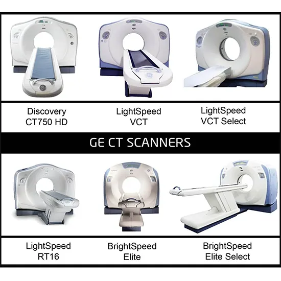
To create cross-sectional images of the body, medical imaging professionals employ CT (Computerised Tomography) machines.
To create cross-sectional images of the body, medical imaging professionals employ CT (Computerised Tomography) machines. CT machines provide precise images of the inside of the body using a combination of X-ray technology and computer software.
The apparatus contains an enormous opening in the shape of a doughnut, known as a gantry, where the patient lies down on a table that glides into the opening. When the patient is inside the gantry, the X-ray tube rotates around them as detectors on the other side of the gantry track the X-rays that travel through their bodies.
There are different types of CT scan machines available, and they differ based on their technology and purpose. Here are some of the most common types of CT scan machines:
Conventional CT scans
One of the first types of CT scanners were conventional CT scanners, commonly referred to as spiral or helical CT scanners. They make a series of 2D images of the body using a focused x-ray beam, which are then merged by a computer to provide a precise 3D image. Key characteristics of traditional CT scanners include:
Rapid diagnosis is essential in emergency situations, making conventional CT scanners excellent. They can scan the complete body in just a few seconds.
High-resolution images can be created by conventional CT scanners, allowing for the detection of tiny lesions or anomalies.
Patients getting traditional CT scans are only permitted to move very little during the process in order to avoid image blur.
Radiation exposure: Older CT scanners release more radiation than modern models, which can raise the risk of developing cancer.
Limited capacity to image specific bodily components, including the lungs and bones, with conventional CT scanners.
Cost-effective: Traditional CT scanners are frequently more affordable than more modern scanners, making them more available to smaller clinics and hospitals.
The majority of hospitals and clinics have access to conventional CT scanners, making them a common tool for detecting a range of medical disorders.
Limited contrast resolution: Due to the limited contrast resolution of traditional CT scanners, it can be challenging to discern between various soft tissue types.
Spiral CT scans
In order to produce accurate 3D images of the body, spiral CT scans, sometimes referred to as helical CT scans, use a sophisticated form of CT scanner that continuously spirals. A spiral CT scanner rotates constantly around the patient, creating a continuous stream of pictures that are merged to form a more detailed 3D image of the body rather than taking discrete "slices" of the body as with traditional CT scans.
Spiral CT scanners may provide images significantly faster than traditional CT scanners and with less radiation exposure to the patient while also producing images of superior quality. They can be used for more precise imaging of the brain and other organs, and are especially helpful for imaging moving organs like the heart and lungs.
Spiral CT scanners come in two primary varieties: single-slice and multi-slice. Multi-slice scanners employ multiple rows of detectors to produce higher-quality images more quickly than single-slice scanners using a single row of detectors to make images.
Cancer, cardiovascular disease, and lung disease are just a few of the illnesses that are frequently diagnosed and monitored with spiral CT scans. Additionally, they are utilised to plan and direct various medical operations like biopsies and radiation therapy.
Dual Energy CT Scanner
Dual-energy CT scanners, sometimes referred to as spectral CT scanners, are a type of CT device that can concurrently collect two sets of data at various energy levels. As a result, the scanner can provide more precise and detailed images by differentiating between various types of tissue depending on their composition and density.
Dual-energy CT scanners have a number of significant features, such as:
Dual-source CT: This type of scanner concurrently collects low- and high-energy data using two x-ray tubes and two detectors. As a result, scanning durations can be shortened and image quality can be increased.
Single-source CT: This type of scanner alternates between low- and high-energy scans using a single x-ray tube and a specific filter. Although it takes longer to gather the required data, it is a more economical choice.
The ability to visualise various substances and bodily features, such as the presence of iodine in contrast agents or the density of bone, is known as spectral imaging.
Virtual non-contrast imaging: This capability can produce a "virtual" non-contrast image using dual-energy data without the requirement for a real non-contrast scan. In addition to increasing patient comfort, this lowers radiation exposure.
Reduction of metal artefact: Dual-energy CT scanners can lessen the artefact brought on by metallic implants, such as those used in joint replacement or dental treatment. This makes it possible to see the surrounding tissue more clearly.
Multi-Slice CT scanner
many rows of detectors: MSCT scanners are equipped with many rows of detectors, allowing them to record more image data with each X-ray tube rotation. As a result, scan times can be shortened and 3D images can be produced.
MSCT scanners can produce images with sub-millimeter resolution, which allows them to capture incredibly minute features within the body.
Lower radiation doses can be used by MSCT scanners compared to older CT scanner models while still delivering images of good quality.
Dynamic scanning: By rapidly obtaining a number of photos throughout a single breath-hold, MSCT scanners can capture images of moving internal body structures like the heart or lungs.
Dual-energy capability: Some MSCT scanners have the ability to produce images with various X-ray energies thanks to their dual-energy capabilities. This may be helpful in identifying specific tissue types, such as calcium deposits or iodine contrast.
Cone-Beam CT Scanner
Technology : Cone-Beam CT is a modified version of conventional CT scanning. The CBCT scanner uses a cone-shaped X-ray beam that spins around the patient instead of a fan-shaped X-ray beam to take several images that are then combined to create a 3D volume of the patient.
Applications : CBCT scanners can be utilised in a variety of settings, including orthopaedics, radiology, and interventional treatments. However, dentistry and maxillofacial imaging is where they are most frequently used.
Benefits : CBCT scanners are superior to other types of CT scanners in a number of ways. They take less time to scan and create 3D images of great quality with minimal radiation exposure.
Cons : Compared to conventional CT scanners, CBCT scanners have a smaller field of vision, which means they can only record images of a small portion of the body. Additionally, CBCT images' resolution might not be as high as that of conventional CT scans.
The Vatech Pax-i3D Green, Planmeca ProMax 3D Mid, and Carestream CS 9300 are a few examples of CBCT scanners.
Photon- Counting CT Scanner
Newer CT scanners called photon-counting CT scanners (PCCT) use cutting-edge detector technology to enhance image quality, cut radiation exposure, and open up new clinical applications.
Photon-counting detectors (PCDs), which can determine the energy of individual x-ray photons, are used in PCCT scanners. Improved contrast resolution is the result of being able to distinguish between tissues of different densities and measuring the x-ray beam with greater precision.
CT imaging could be completely changed by PCCT technology, especially in the areas of cardiology, cancer, and neurology. PCCT can assist in the early detection of tiny lesions and assist in monitoring the development of disease because it has the capacity to precisely differentiate between various types of tissues.
By more accurately defining tumour boundaries and sparing healthy tissue, PCCT can significantly increase the precision of radiation therapy planning.
Portable CT Scanner
Instead of having the patient transported to the machine, portable CT scanners are compact devices that may be transported to the patient's location. They are frequently employed in emergency and critical care settings when quick and simple imaging is required, as well as in distant or resource-constrained locations where a stationary CT scanner may not be available.
Some essential characteristics of mobile CT scanners include
Lightweight construction : Portable CT scanners are made to be portable and compact, making it simple to move them around and set them up in different areas.
Battery-operated : Since many portable CT scanners come with built-in batteries, they can be used in locations without access to an electrical outlet.
Rapid imaging is a service that portable CT scanners can offer, with scan times varying from a few seconds to a few minutes.
The head, chest, abdomen, and extremities are just a few of the body areas that can be imaged using portable CT scanners due to their versatility.
Low radiation exposure : Some mobile CT scanners feature cutting-edge imaging technologies that reduce radiation exposure, making them perfect for usage with children and other radiation-sensitive groups.


