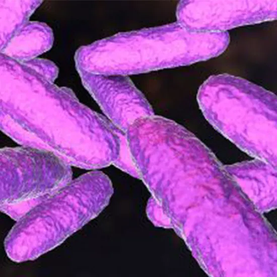
The gram-negative bacteria Klebsiella granulomatis is the cause of the STI granuloma inguinale, often known as donovanosis, which is a sexually transmitted infection (STI). Although it can affect other parts of the body, this...
The gram-negative bacteria Klebsiella granulomatis is the cause of the STI granuloma inguinale, often known as donovanosis, which is a sexually transmitted infection (STI). Although it can affect other parts of the body, this illness typically affects the skin and mucous membranes of the vaginal area.
The condition is characterized by the development of painless, beefy-red, granulomatous ulcers that can bleed and become infected in the vaginal area. The surrounding tissues may be damaged and enlarged by the ulcers, which can result in deformity, chronic ulcers, and, in severe cases, elephantiasis.
K. granulomatis is mainly found in tropical and subtropical areas, and it can spread through non-sexual contact with infected objects in addition to sexual contact. The ulcers are often visually inspected to make the diagnosis, which is then confirmed by laboratory studies. A course of antibiotics such as ciprofloxacin, azithromycin, or doxycycline is typically prescribed as part of the treatment.
Donovanosis can be prevented by wearing condoms, having safe intercourse, and avoiding contact with sick people or their contaminated objects. To avoid problems and stop the spread of the disease, early diagnosis and treatment of the condition are crucial.
Klebsiella granulomatis A century-long story of research and discovery
Charles Donovan, a British doctor, originally described Klebsiella granulomatis in 1905. Donovan named the bacteria "Donovan bodies" after himself after noticing the distinctive intracellular inclusions in tissue samples from patients with vaginal ulcers.
Initially, it was believed that K. granulomatous-related illnesses could only be found in particular parts of the world, such as Southeast Asia, Africa, and India. However, instances were subsequently documented in Australia, Europe, and the Americas, among other regions of the globe.
Granulomatous ulcers in the vaginal area were the disease's distinctive symptom, and the term "granuloma inguinale" was first used to describe it in the 1910s. In the past, the condition was also known as "venereal granuloma" and "pudendal ulcer."
As laboratory methods have advanced and more powerful antibiotics have been accessible, the diagnosis and management of donovanosis have changed throughout time. However, donovanosis continues to be a serious public health issue, particularly in underdeveloped nations with little access to safe sex education and treatment.
A closer look at Klebsiella granulomatis: Understanding its fascinating morphology
The key characteristics of Klebsiella granulomatis' morphology are as follows:
Gram-negative: K. granulomatis exhibits gram-negative staining, in which case it loses the crystal violet stain during the Gramme staining process and exhibits a reddish or pinkish appearance upon counterstaining.
Shape:K. granulomatis is a short, plump rod-shaped bacterium that is usually 1-2 micrometers long and 0.5-0.8 micrometers wide.
Non-motile: K. granulomatis is not able to move independently because it lacks flagella.
Capsule:K. granulomatis is encapsulated, which means that a polysaccharide capsule surrounds it and serves to shield it from the host's defenses.
Intracellular Inclusion:Donovan bodies, which are generally round or oval and measure 1-2 micrometers in diameter, and include bacterial clusters around by a halo-like structure, are distinctive intracellular inclusions that K. granulomatis produces.
Mucoid Colonies:K. granulomatis colonies formed on culture media often have a mucoid appearance and are tiny, smooth, and convex. They can be challenging to recognize from other non-lactose fermenting bacteria due to their typical grayish-white color.
The identification and diagnosis of K. granulomatis in the laboratory depend on these morphological characteristics.
Klebsiella granulomatis life cycle: From colonization to tissue damage
Since Klebsiella granulomatis is a bacterium that reproduces by binary fission, it does not have a regular life cycle. It doesn't go through a complicated life cycle that involves sexual reproduction or spore development, as a result.
Instead, direct skin-to-skin contact or sexual contact with an infected person is how the bacteria enter the body. Once inside the body, it infiltrates macrophages and reproduces there, producing distinctive intracellular inclusions known as Donovan bodies. The bacteria may withstand periods inside macrophages, eluding the human immune system and leading to persistent infection.
The main stages in the life cycle of this disease include the transfer of K. granulomatis from an infected person to a vulnerable host, as well as the invasion and multiplication of the bacterium within host cells. To halt the disease's course and stop its spread, early identification, and adequate antibiotic therapy are essential.
Klebsiella granulomatis infection: An overlooked but emerging threat to sexual health
The key characteristics of donovanosis, commonly known as a Klebsiella granulomatis infection, are as follows:
Transmission
Direct skin-to-skin contact or sexual activity with an infected person are the two main ways in which the illness is spread.
Incubation Period
Donovanosis normally has an incubation period of 1 to 12 weeks or an average of 50 days.
Clinical Presentation
Donovanosis presents clinically as a painless papule or nodule that can develop into a superficial ulcer with a beefy red look. The genitalia, perineum, or anus are the most common sites for ulcers, although they can also develop in other regions of the body. The ulcers can be solitary or many.
Tissue destruction
If the infection is not treated, it may spread to Tissue Culture and Sensitivity other tissues and result in significant tissue loss, which might leave scars and cause deformity.
Geographical distribution
Tropical and subtropical locales, including sections of Africa, India, Papua New Guinea, and some regions of Central and South America, are where donovanosis is most commonly seen.
These distinguishing characteristics of Klebsiella granulomatis infection emphasize the significance of early detection and adequate treatment to stop the disease's progression and its implications.
Klebsiella granulomatis diagnosis: From microscopy to molecular methods
Donovanosis, commonly known as an infection by Klebsiella granulomatis, is diagnosed using a combination of clinical assessment and laboratory investigations. Here are a few typical diagnostic techniques:
Clinical assessment
The first step in the diagnosis of donovanosis is frequently a thorough physical examination of the afflicted region by a healthcare professional. The characteristic lesion of donovanosis is a raised, beefy-red ulcer that is painless to the touch and may bleed readily.
Microscopy
To check for the existence of distinctive intracellular Donovan bodies, a tissue sample or swab from the lesion is taken and examined under a microscope. Within host cells, the bacteria are grouped in oval-shaped structures called Donovan bodies.
Swab
To isolate, confirm and confirm the diagnosis of donovanosis, the biopsy or swab sample may also be cultivated in a specialized laboratory. This procedure could need specialized lab equipment and might be time-consuming.
Polymerase chain reaction (PCR )
Using the polymerase chain reaction (PCR), it is possible to identify the bacterial DNA in clinical samples. This technique may quickly diagnose donovanosis and is very sensitive and specific.
Serological testing
Blood tests that look for antibodies to K. granulomatis are not frequently used in clinical practice but may help confirmtrackosis or tracking therapy effectiveness.
It is significant to highlight that donovanosis can be difficult to diagnose because of its rarity and vague clinical symptoms, and it may be mistaken for other genital ulcers such as syphilis or chancroid. For appropriate diagnosis and treatment, healthcare professionals must thus maintain a high level of clinical suspicion and employ a variety of diagnostic techniques.
Klebsiella granulomatis treatment: A global health priority that requires action
Antibiotics
Antibiotics are used to treat Klebsiella granulomatis infection, commonly known as donovanosis, to get the bacterium out of the body. The selection of antibiotics and length of therapy is based on the infection's severity and the patient's general condition. To treat donovanosis, the following antibiotics are frequently used:
Azithromycin
Mild to moderate cases of donovanosis can be successfully treated with a single dosage of 1 g of azithromycin.
Doxycycline
For more serious or complex instances of donovanosis, a course of doxycycline for 21 days is the best therapy.
Tetracycline
Patients who cannot take tetracycline antibiotics may use ciprofloxacin as an alternative to doxycycline.
Trimethoprim-sulfamethoxazole
It is an additional treatment option for donovanosis in people who are unable to take tetracycline medications.
Supportive therapy
Supportive therapies, such as painkillers and anti-inflammatory drugs, could also be required to treat the donovanosis's symptoms, such as inflammation and discomfort. To stop future transmission, patients should be urged to refrain from sexual activity until the illness has cleared up.
It is essential to remember that fast diagnosis and treatment are crucial for halting the spread of infection and avoiding consequences including substantial tissue damage and scarring. To ensure that the infection is adequately treated and that there is no recurrence, patients should be constantly watched while undergoing therapy.
Don't let Klebsiella granulomatis win : Join the fight and take control of your health.


