Gnathostoma spinigerum and Gnathostoma hispidum are parasitic nematodes that belong to the household Gnathostomatidae. Both species are many times referred to as Gnathostoma worms and are recognised to infect a large variety...
Gnathostoma spinigerum and Gnathostoma hispidum are parasitic nematodes that belong to the household Gnathostomatidae. Both species are many times referred to as Gnathostoma worms and are recognised to infect a large variety of mammalian and fish hosts.
Gnathostoma spinigerum is the most frequent species observed in human beings and is regarded to purpose gnathostomiasis, a parasitic contamination that can lead to extreme signs such as pores and skin lesions, migratory swelling, and neurological damage.
The contamination is generally obtained with the aid of eating uncooked or undercooked fish, eels, frogs, or birds that include infective larvae. Gnathostoma hispidum, on the other hand, is mainly determined in wild carnivores such as foxes and wildcats and is much less usually mentioned in humans.
What are Gnathostoma spinigerum and Gnathostoma hispidum Infections?
Gnathostoma spinigerum and Gnathostoma hispidum infections are parasitic ailments prompted by the way of the respective nematode species, Gnathostoma spinigerum and Gnathostoma hispidum.
Both infections are jointly referred to as gnathostomiasis and can have an effect on people and a range of different animals, which includes cats, dogs, pigs, and wild animals. In humans, gnathostomiasis is usually received using ingesting uncooked or undercooked fish or different seafood that is contaminated with the larvae of the worm.
The larvae can additionally penetrate the pores and skin of people when they come into contact with contaminated water or soil.
Explore the Epidemiology of Gnathostoma spinigerum and Gnathostoma hispidum:
Gnathostoma spinigerum and Gnathostoma hispidum infections are endemic in countless international locations in Asia, consisting of Thailand, Japan, China, and the Philippines.
However, instances have additionally been mentioned in different components of the world, which include Central and South America, Africa, and even some components of Europe.
The incidence of gnathostomiasis varies relying on the area and populace studied. In endemic areas, the incidence of contamination can be as excessive as 10-20% in some communities.
The best incidence of contamination happens in rural and fishing communities where the consumption of uncooked or undercooked fish is common.
Gnathostomiasis can affect human beings of all ages, however, it is more frequent in adults than in children. Males are more often affected than females, and the contamination is extra frequent in humans who devour uncooked or undercooked fish or seafood.
In animals, gnathostomiasis is additionally frequent in home and wild cats, dogs, and different carnivorous animals. The transmission of the ailment between animals and human beings is possible, making gnathostomiasis a zoonotic disease.
Gnathostoma hispidum infections are fairly uncommon in human beings and are typically observed in areas where wild carnivorous animals, such as foxes and wildcats, are prevalent.
The transmission of G. hispidum to people is no longer nicely understood, however, it is thought to happen via ingestion of contaminated intermediate hosts or contaminated water.
Learn about the Pathophysiology of Gnathostoma spinigerum and Gnathostoma hispidum:
Gnathostoma spinigerum and Gnathostoma hispidum infections end in a parasitic ailment known as gnathostomiasis, which is prompted by way of the nematodes of the Gnathostoma genus.
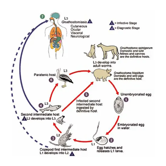
The pathophysiology of these infections is complicated and includes various stages. The existence cycle of Gnathostoma includes a couple of hosts, along with freshwater crustaceans, fish, amphibians, reptiles, birds, and mammals.
The grownup worms stay in the belly of definitive hosts, such as cats and dogs, and produce eggs that are excreted in the host's faeces.
These eggs hatch in water, releasing free-swimming larvae that infect freshwater crustaceans, which are then fed on by way of fish or different intermediate hosts.
When human beings devour uncooked or undercooked fish or different intermediate hosts that comprise infective larvae, the larvae penetrate the intestinal wall and migrate to a range of tissues and organs.
They might also motivate harm as they move, mainly to irritation and the formation of nodules. The larvae can additionally migrate to the talent and spinal cord, mainly to neurological symptoms.
The pathophysiology of gnathostomiasis is often due to the host's immune response to the migrating larvae.
The larvae have a special capacity to steer clear of the host's immune device and cause inflammation, mainly to signs such as itching, migratory pores and skin swellings, and belly pain.
The host's immune device can additionally harm surrounding tissues and organs in the course of the inflammatory response.
Severe instances of gnathostomiasis can lead to life-threatening problems such as eosinophilic meningitis, encephalitis, and pulmonary eosinophilia.
The genuine mechanisms main to these problems are no longer entirely understood, however, it is an idea to be due to the host's immune response to the migrating larvae.
The pathophysiology of Gnathostoma hispidum infections is comparable to that of Gnathostoma spinigerum infections, however, the contamination is much less frequent and commonly milder in humans.
Signs and Symptoms of Gnathostoma spinigerum and Gnathostoma hispidum Infection
The symptoms and signs of Gnathostoma spinigerum and Gnathostoma hispidum infections, Urine Routine at the same time regarded as gnathostomiasis, can differ depending on the region of the larvae in the physique and the severity of the infection.
Some human beings contaminated with Gnathostoma may additionally be asymptomatic or have solely slight symptoms, whilst others can also improve extreme and life-threatening complications.
Common symptoms and signs and symptoms of gnathostomiasis include:
Migratory pores and skin swellings or lumps
These are the most common and attributed symptoms of gnathostomiasis. The swellings may additionally be painful, and itchy, and pass around the physique as the larvae migrate.
Subcutaneous nodules
These are firm, raised lumps that may additionally enhance beneath the pores and skin and are prompted using the larvae migrating via the subcutaneous tissue.
Fever and flu-like symptoms
Some humans might also increase fever, headache, muscle aches, and different flu-like symptoms.
Gastrointestinal symptoms
These might also encompass belly pain, nausea, vomiting, and diarrhoea.
Neurological symptoms
In extreme cases, the larvae can migrate to the talent and spinal cord, inflicting neurological signs such as meningitis, encephalitis, seizures, paralysis, and even coma.
The signs of Gnathostoma hispidum infections are comparable to those of Gnathostoma spinigerum infections, however, they are commonly milder and much less common. It is necessary to notice that the symptoms and signs and symptoms of gnathostomiasis can be non-specific and overlap with those of different diseases.
Therefore, laboratory assessments and imaging research are commonly wished to verify the diagnosis.
Diagnosis of Gnathostoma spinigerum and Gnathostoma hispidum Infections
The analysis of Gnathostoma spinigerum and Gnathostoma hispidum infections, jointly regarded as gnathostomiasis, can be difficult due to the fact the signs can be non-specific and overlap with those of different diseases.
Therefore, laboratory exams and imaging research are commonly wanted to verify the diagnosis.
The following diagnostic assessments can also be used for the detection of Gnathostoma infection:
Blood tests
An entire blood count number (CBC) may additionally exhibit an enlargement in eosinophil count, which is a frequent characteristic of parasitic infections.
Serological tests
Enzyme-linked immunosorbent assay (ELISA) and Western blot are serological exams that can notice antibodies in opposition to Gnathostoma in the blood.
Imaging studies
X-rays, ultrasound, computed tomography (CT) scans, and magnetic resonance imaging (MRI) can also be used to observe subcutaneous nodules, or larvae in tissues and organs, such as the brain, spinal cord, and eyes.
Biopsy
A biopsy of the affected tissue or nodule can also disclose the presence of Gnathostoma larvae.
Stool examination
The presence of Gnathostoma eggs in the stool might also advocate the diagnosis, however, this is now not a dependable approach due to the fact the eggs are regularly now not excreted in the stool.
It is necessary to say that the analysis of gnathostomiasis can also be challenging, and it is regularly based totally on an aggregate of scientific presentations, laboratory tests, and imaging studies.
Therefore, it is vital to seek advice from a scientific expert skilled in diagnosing and treating parasitic infections.
Complications of Gnathostoma spinigerum and Gnathostoma hispidum Infections
Gnathostoma spinigerum and Gnathostoma hispidum infections, mutually recognized as gnathostomiasis, can cause a range of complications, in particular, if the larvae migrate to different components of the body.
The severity of the issues relies upon the place and range of larvae, the period of the infection, and the immune fame of the contaminated person.
Some of the viable problems of gnathostomiasis include:
Meningitis and encephalitis
If the larvae migrate to the central frightened system, they can cause infection of the Genius and spinal cord, mainly to neurological signs such as headache, confusion, seizures, and paralysis.
Eye involvement
If the larvae migrate to the eyes, they can motivate irritation of the eye, leading to signs and symptoms such as redness, pain, and imaginative and prescient loss.
Respiratory symptoms
In uncommon cases, the larvae can migrate to the lungs, inflicting signs and symptoms such as cough, shortness of breath, and chest pain.
Gastrointestinal complications
If the larvae migrate to the gastrointestinal tract, they can cause irritation and ulceration, mainly signs such as belly pain, nausea, vomiting, and diarrhoea.
Allergic reactions
Some humans might also boost allergic reactions to the larvae or their excretions, mainly to signs such as hives, itching, and anaphylaxis.
Death
In extreme cases, such as when the larvae migrate to the Genius or spinal cord, gnathostomiasis can be fatal.
It is vital to be aware that now not everybody contaminated with Gnathostoma will improve complications, and the severity of the contamination can range greatly.
However, it is necessary to try to find clinical interest if you suspect you might also have gnathostomiasis, as early analysis and remedy can assist stop complications.
Treatment of Gnathostoma spinigerum and Gnathostoma hispidum Infection
The cure of Gnathostoma spinigerum and Gnathostoma hispidum infections, mutually recognised as gnathostomiasis, normally includes a mixture of medicines and supportive care.
The following redress can also be used for gnathostomiasis:
Anthelmintic medications
Albendazole and ivermectin are the most generally used medicinal drugs for the remedy of gnathostomiasis. These medicines work by way of killing the larvae and stopping them from migrating to different components of the body. The treatment period can also range relying on the severity of the infection.
Pain management
Pain administration may additionally be integral to relieving signs such as headache, stomach pain, and joint pain.
Non-steroidal anti-inflammatory tablets (NSAIDs) or different ache medicinal drugs can also be prescribed.
Steroids
In instances of extreme inflammation, such as when the larvae migrate to the Genius or spinal cord, steroids may additionally be prescribed to decrease infection and stop complications.
Surgery
In uncommon cases, a surgical procedure can also be imperative to put off the larvae or to deal with issues such as abscesses. It is essential to be aware that early prognosis and remedy are key to stopping issues from gnathostomiasis.
Therefore, if you suspect you may additionally have gnathostomiasis, try to find scientific interest as quickly as possible.
Additionally, it is vital to take steps to forestall publicity to the larvae, such as fending off the consumption of undercooked fish or eels and keeping off consuming water from untreated sources.
Exploring the mysteries of Gnathostoma Spinigerum and Gnathostoma Hispidum: The Silent Invaders with Deadly Impacts


