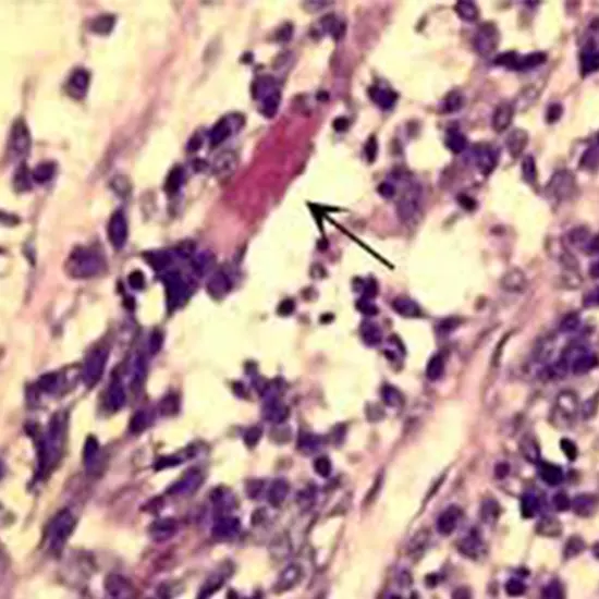
Rhabdomyosarcoma (RMS) may be a profoundly forceful shape of cancer that arises from mesenchymal cells that have fizzled to completely separate into myocytes of skeletal muscle. Cells of the tumor are recognized as...
Rhabdomyosarcoma (RMS) may be a profoundly forceful shape of cancer that arises from mesenchymal cells that have fizzled to completely separate into myocytes of skeletal muscle. Cells of the tumor are recognized as rhabdomyoblasts.
There are four subtypes
- Embryonal rhabdomyosarcoma
- Alveolar rhabdomyosarcoma
- Pleomorphic rhabdomyosarcoma
Spindle cell/sclerosing rhabdomyosarcoma. Embryonal and alveolar are the most common, and these sorts are the foremost common delicate tissue sarcomas of childhood and youth. The pleomorphic sort is ordinarily found in grown-ups.
It is by and large considered to be an infection of childhood, as the endless larger part of cases happens in those under the age of 18. It is commonly portrayed as one of the small-blue-round-cell tumors of childhood due to its appearance on an H&E recolor. Despite being generally uncommon, it accounts for roughly 40% of all recorded delicate tissue sarcomas.
RMS can happen in any delicate tissue location within the body but is fundamentally found within the head, neck, circle, genitourinary tract, private parts, and limits. There are no clear chance components, but the malady has been related to a few inherent abnormalities. Signs and side effects change concurring to tumor location, and the forecast is closely tied to the area of the primary tumor. Common locales of metastasis incorporate the lungs, bone marrow, and bones. There are numerous classification frameworks for RMS and an assortment of characterized histological sorts. Embryonal rhabdomyosarcoma is the foremost common sort and comprises around 60% of cases.
Results change significantly, with 5-year survival rates between 35% and 95 pending on the type of RMS included, so a clear conclusion is basic for viable treatment and administration. Precise and fast determination is regularly troublesome due to the heterogeneity of RMS tumors and a need for solid hereditary markers of the malady, even though later inquiries by UVA Wellbeing analysts found “multiple lines of prove supporting [the quality] AVIL is a capable driver for both major sorts of rhabdomyosarcoma,” concurring to analyst Hui Li, Ph.D., of the College of Virginia School of Medicine's Office of Pathology and UVA Cancer Center. In a logical paper sketching out the discoveries, he and his colleagues portray rhabdomyosarcoma as “addicted” to the gene's abundance movement. They eventually named AVIL a “bona fide oncogene” for rhabdomyosarcoma.
Treatment ordinarily includes a combination of surgery, chemotherapy, and radiation. Sixty percent to 70% of recently analyzed patients with nonmetastatic illness can be cured utilizing this combined approach to treatment. Despite forceful multimodality treatment, less than 20% of patients with metastatic RMS can be cured of their infection.
Types
Given the trouble in diagnosing rhabdomyosarcoma, conclusive classification of subtypes has demonstrated troublesome. As a result, classification frameworks change by the organization. In any case, rhabdomyosarcoma within the 2020 WHO classification is recorded as four histological subtypes: embryonal, alveolar, pleomorphic, and shaft cell/sclerosing.
Embryonal
Embryonal rhabdomyosarcoma (ERMS) is the foremost common histological variation, comprising roughly 60–70% of childhood cases. It is most common in children 0–4 a long time ago, with the greatest detailed rate of 4 cases per 1 million children. ERMS is characterized by spindle-shaped cells with a stromal-rich appearance, and the morphology is comparative to the making muscle cells of a 6- to 8-week-old developing life. Tumors frequently show within the head and neck as well as the genitourinary tract.
Embryonal subtype
Botryoid rhabdomyosarcoma is nearly continuously found in mucosal-lined organs counting the vagina, bladder, and nasopharynx (although introduction within the nasopharynx ordinarily influences more seasoned children). It frequently presents in newborn children more youthful than a year old, as a circular, grape-like mass on the influenced organ. Histologically, cells of the botryoid variation are characterized by a thick tumor layer beneath an epithelium (cambium layer). This subtype includes a great forecast.
Botryoid rhabdomyosarcoma is additionally some of the time displayed in grown-up ladies, found within the cervix or uterus.
Alveolar
Alveolar rhabdomyosarcoma (ARMS) is the moment most common sort. ARMS comprises around 20–25% of RMS-related tumors, and it is similarly conveyed among all age bunches with a rate of approximately 1 case per 1 million individuals aged 19. For this reason, it is the foremost common shape of RMS watched by youthful grown-ups and young people, who are less inclined to the embryonal variation. This sort of RMS is characterized by densely-packed, circular cells that organize around spaces comparable in shape to respiratory alveoli, even though variations have been found without these characteristic alveolar spacings. ARMS tends to create more often within the limits, trunk, and peritoneum. It is additionally ordinarily more forceful than ERMS.
Pleomorphic
Pleomorphic rhabdomyosarcoma (undifferentiated rhabdomyosarcoma), too known as anaplastic rhabdomyosarcoma, is characterized by the nearness of pleomorphic cells with huge, lobate hyperchromatic cores and multipolar mitotic figures. These tumors show tall heterogeneity and greatly destitute separation. The pleomorphic cells may be diffuse or localized, with the diffuse variety relating to a more regrettable guess. It happens most frequently in grown-ups, once in a while in children, and is regularly found within the extremities. Due to the need for perceivable partition among cancers of this sort, clinicians will frequently name undiscovered sarcomas with small to no recognizable highlights as anaplastic RMS. It is the foremost forceful sort of RMS, and will regularly require serious treatment.
Spindle cell/sclerosing
Axle cell/sclerosing rhabdomyosarcoma is an included subtype recorded within the 2020 WHO classification of delicate tissue sarcomas.
This subtype is exceptionally comparative to that of leiomyosarcoma (cancer of the smooth muscle tissue), and it includes a fascicular, spindled, and leiomyomatosis growth design with eminent rhabdomyoblastic separation. It happens most commonly within the para testicular locale, and the guess for this specific frame of RMS is amazing with a detailed five-year survival rate of 95%. The sclerosing perspective of this subtype has hyaline sclerosis and neurovascular development.
Numerous classification frameworks have been proposed for directing administration and treatment, and the foremost later and broadly utilized classification framework is the "Universal Classification of Rhabdomyosarcoma" or ICR. It was made by the IRSG in 1995 after their arrangement of four multi-institutional trials pointed at considering the introduction, histology, study of disease transmission, and treatment of RMS (IRSG I–IV). The ICR framework is based on prognostic pointers recognized in IRSG I–IV. Pleomorphic rhabdomyosarcoma ordinarily happens in grown-ups instead of children and is hence not included in this framework.
Signs and Symptoms
RMS can happen in nearly any soft-tissue location within the body; the foremost common essential destinations are genitourinary (24%), para meningeal (16%), limit (19%), circle (9%), other head and neck (10%), and different other destinations (22%).
RMS regularly presents as a mass, but signs and indications can shift broadly depending on the location of the essential tumor. Genitourinary tumors may display hematuria, urinary tract obstacle, and/or a scrotal or vaginal mass.
Tumors that emerge within the retroperitoneum and mediastinum can end up very huge sometime recently creating signs and indications.
Parameningeal tumors may display cranial nerve brokenness, side effects of sinusitis, ear release, cerebral pains, and facial torment. Orbital tumors frequently show orbital swelling and proptosis. Limit tumors for the most part show as a rapidly extending, firm mass within the pertinent tissue.
Cancer's predominance within the head confronts, and the neck will regularly permit prior signs of the illness basically due to the self-evident nature of tumors in these locations. Despite the shifting introduction and regularly forceful nature of the malady, RMS has the potential to be analyzed and treated early.
The fourth IRSG review found that 23% of patients were analyzed in time for total resection of their cancer, and 15% had resection with as it were negligible leftovers of the ailing cells.
Risk Factors
Rhabdomyosarcoma is troublesome to analyze. Chance components that increment the probability of this cancer incorporate acquired clutter such as Li-Fraumeni disorder, Neurofibromatosis sort 1, Beckwith-Wiedemann disorder, Costello disorder, Noonan disorder, and DICER1 disorder.
Diagnosis
Rhabdomyosarcoma is regularly troublesome to analyze due to its likenesses to other cancers and changing levels of separation. It is freely classified as one of the small-blue-round-cell tumors due to its appearance on an H&E recolor.
Other cancers that share this classification incorporate neuroblastoma, Ewing sarcoma, and lymphoma, and a determination of RMS requires a sure end of these morphologically comparative diseases. The characterizing symptomatic characteristic of RMS is an affirmation of threatening skeletal muscle separation with myogenesis (displaying as a stout, pink cytoplasm) beneath light microscopy. Cross striations may or may not be displayed.
Exact determination is more often than not finished through immunohistochemical recoloring for muscle-specific proteins such as myogenin, muscle-specific actin, desmin, D-myosin, and myoD1. Myogenin, in specific, has appeared to be exceedingly particular to RMS, although the demonstrative noteworthiness of each protein marker may change depending on the sort and area of the dangerous cells.
The alveolar sort of RMS tends to have more grounded muscle-specific protein recoloring. Electron microscopy may too help in the conclusion, with the nearness of actin and myosin or Z groups indicating a positive conclusion of RMS. Classification into sorts and subtypes is finished through assisted investigation of cellular morphology (alveolar spacings, nearness of cambium layer, aneuploidy, etc.) as well as hereditary sequencing of tumor cells.
A few hereditary markers, such as the PAX3-FKHR combination quality expression in alveolar RMS, can help in the conclusion. Open biopsy is more often than not required to get adequate tissue for precise determination. All discoveries must be considered in the setting, as no one character could be a conclusive pointer for RMS.
Staging
After conclusion and histopathological investigation, different imaging procedures may be utilized, counting MRI, ultrasound, and a bone check in arrange to decide the degree of nearby attack and any metastasis. Encouraging investigational strategies may be vital depending on tumor locales. A para meningeal introduction of RMS will regularly require a lumbar cut to run the show-out metastasis to the meninges. A para testicular introduction will frequently require a stomach CT to run the show out nearby lymph hub inclusion, and so on. Results are emphatically tied to the degree of the illness, and its early mapping is critical for treatment arranging.
The current organizing framework for rhabdomyosarcoma is abnormal relative to most cancers. It utilizes an altered TNM (tumor-nodes-metastasis) framework initially created by the IRSG. This framework accounts for tumor measure (> or <5 cm), lymph hub inclusion, tumor location, and nearness of metastasis. It grades on a scale of 1 to 4 based on these criteria. In expansion, patients are sorted by clinical gather (from the clinical bunches from the IRS considers) based on the victory of their to begin with surgical resection. The current Children's Oncology Gather conventions for the treatment of RMS categorize patients into one of four hazard categories based on tumor review and clinical bunch, and these hazard categories have appeared to be exceedingly prescient of the results.
Treatment
Treatment of rhabdomyosarcoma may be multidisciplinary hone including the utilization of surgery, chemotherapy, radiation, and conceivably immunotherapy. Surgery is by and large the primary step in a combined helpful approach. Respectability shifts depending on tumor location, and RMS regularly presents in locales that do not permit full surgical resection without critical dreariness and misfortune of work. Less than 20% of RMS tumors are completely resected with negative edges. Rhabdomyosarcomas are exceedingly chemosensitive, with roughly 80% of cases reacting to chemotherapy. In reality, multi-agent chemotherapy is shown for all patients with rhabdomyosarcoma. Sometime recently the utilization of adjuvant and neoadjuvant treatment including chemotherapeutic operators, treatment exclusively by surgical implies had a survival rate of <20%. Present-day survival rates with adjuvant treatment are roughly 60–70%.
There are two fundamental strategies of chemotherapy treatment for RMS. There's the VAC regimen, comprising vincristine, actinomycin D, and cyclophosphamide, and the IVA regimen, comprising ifosfamide, vincristine, and actinomycin D. These drugs are managed in 9–15 cycles depending on the arranging of the infection and other treatments utilized. Other sedate and treatment combinations may moreover show an additional advantage. Expansion of doxorubicin and cisplatin to the VAC regimen appeared to extend survival rates of patients with alveolar-type, early-stage RMS in IRS think about III, and this same expansion moved forward survival rates and multiplied bladder rescue rates in patients with organized III RMS of the bladder. In children and youthful grown-ups with arranged IV metastatic rhabdomyosarcoma, a Cochrane survey has found no proof to back the utilization of high-dose chemotherapy as a standard treatment.


