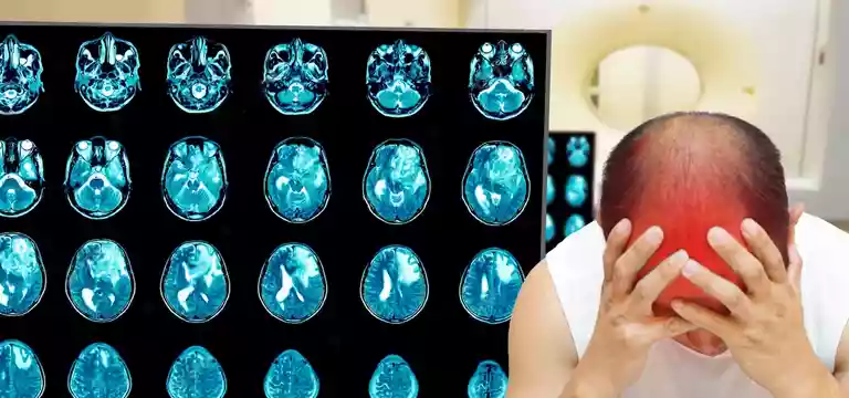
Before we explore the MRI brain epilepsy protocol, we need to understand what is an MRI or magnetic resonance imaging. It is an imaging diagnostic tool based on the principles of using magnetic fields and radio waves to...
Introduction
Before we explore the MRI brain epilepsy protocol, we need to understand what is an MRI or magnetic resonance imaging. It is an imaging diagnostic tool based on the principles of using magnetic fields and radio waves to capture accurate and detailed images of the human body. It does not require the use of X-rays to click the images. So it is helpful for a patient with epilepsy, as an MRI scan may help determine the underlying cause.
MRI protocol for epilepsy
It is a specialized combination of MRI sequences created to boost the accuracy of detecting structural abnormalities associated with seizure disorders. With the help of these specific sequences, such as high-resolution T1-weighted imaging and fluid-attenuated inversion recovery (FLAIR), the sensitivity and specificity of MRI immensely improve, which helps in detecting conditions like mesial temporal sclerosis and malformation of cortical development.
It is important to note that MRI, especially 3 Tesla MRI, is the most preferred choice for investigating epilepsy. The MRI 3T is characterized by higher magnetic field strength and has more capability to capture images of the brain with precision.
So, the MRI brain epilepsy tool is a comprehensive imaging technique playing a crucial role in the diagnosis and management of epilepsy, augmenting the process of understanding the underlying structural factors contributing to seizure activity. It comes as a great help for doctors in formulating personalized treatment plans for people grappling with epilepsy.
So, is an MRI scan safe for a person with epilepsy?
Yes, it is considered safe for people with epilepsy. The MRI scan poses no threat to an individual suffering from epilepsy and for that matter to any patient. But, the appropriate safety measures have to be followed.
Even many heart surgery patients and people who have medical devices implanted are safely examined with MRI:
However, discuss and divulge every detail about any implants to your doctor beforehand. Talk to the physician about any of the following devices, if you have them in your body:
- Surgical clips or sutures
- Staples
- Artificial joints
- Disconnected medication pumps
- Most heart valve replacements
- Vena cava filters
- Brain shunt tubes for hydrocephalus, etc.
In certain cases and conditions, an MRI is not advisable. So talk to your healthcare provider if you have any of the following conditions:
Heart pacemaker
If a patient has a cerebral aneurysm clip (a metal clip on a blood vessel in the brain), then it is not safe to undergo an MRI. However, the latest clips are made of titanium and are safe for an MRI. The doctor is the best judge in this case, so discuss it beforehand.
Pregnancy
Talk about your pregnancy or if you may have any doubts about being pregnant. The doctor will take a decision based on the individual case.
Also discuss any implanted insulin pump, narcotics pump, or implanted nerve stimulators ("TENS") for back pain with the physician.
If you have metal in the eye or eye socket.
When there is a Cochlear (ear) implant for hearing impairment
In the case of older implanted spine stabilization rods (New ones are titanium and safe), talk to your doctor
Any severe lung disease
If you are unable to lie on your back for 30 to 60 minutes
If you have Claustrophobia (fear of closed or narrow spaces). If you have this fear, the doctor will give you a sedative to assuage the anxiety so that you remain calm and relaxed during the MRI scan. But, for all these, there have to be prior arrangements.
How long does it take to complete the MRI scan?
Typically, an MRI exam takes about close to 30 minutes. But, the duration may get extended when the doctor looks for any additional or specialized studies. Throughout the scan, the technician tries to capture several dozen images so that a comprehensive evaluation can be done. These images are very crucial in offering many insights about the area being examined, helping doctors in the diagnosis and treatment planning process. More than the duration of the MRI exam, what is important is acquiring high-quality images for proper assessment of the condition.
Understanding the MRI procedure
One should leave any personal items such as watches, wallets, credit cards with magnetic strips, and jewelry at home or remove them before entering the MRI room. In facilities, they have secure lockers for keeping the personal belongings of the patients. Before the scan, the patient will need to change into a loose-fitting gown, which is provided by the facility.
What to expect during the MRI scan
Once the MRI scan begins, there will be a lot of sounds. The patient will hear a muffled thumping sound, which comes from the equipment, and it will continue for several minutes. The patient will not feel anything apart from the sound, like any unusual sensations during the scanning process. Some patients may have to be administered a contrast agent called gadolinium. It is injected to increase the visibility of certain structures in the scan images.
If the patient has any doubts or reservations regarding the scan, he or she should immediately talk to the technician present or the doctor in advance. They are there to conduct the process and see that it goes on smoothly with optimum findings from the images.
Once the MRI Exam is over, the patient can resume daily activities like normal. However, if he or she was given a sedative for any anxiety, then driving home would not be a safe option. In that case, someone should drive the person home, whether a friend or family.
The results of the MRI will be provided to the concerned doctor within 24 hours, on weekdays. Thereafter, the physician will take up the report and discuss it in detail with the patient. In most cases, the patient will be given a CD containing the study.
MRI Brain Epilepsy Protocol Price
Many patients may have a concern regarding the price of an MRI because it is costly, being a sophisticated diagnostic technique. If you are looking for an MRI brain epilepsy protocol price, then it is important to know that it is different in different scenarios. The cost of an MRI can vary due to the particular location of the facility, the standard of the facility, the use of a contrast dye, special and extra screenings needed for the particular scan, and many others. But, despite the cost, one must not avoid getting the MRI for the sake of one’s health and safety.
Conclusion
MRI brain epilepsy protocol can help doctors in the diagnosis, studying the conditions, and management of epilepsy in an individual.
We have also seen that the MRI protocol for epilepsy is a special combination of MRI sequences intended to enhance the precision of finding out structural abnormalities related to epileptic seizures.
FAQs
Can an MRI detect all types of epilepsies?
No, it may not. It can help the doctor in comprehending the structural causes of the seizures. But, for patients where there is no structural cause behind the seizure, the MRI scan will come as normal.
Is MRI better than EEG?
EEG is more versatile in offering information about the brain, whereas an MRI is not capable of doing that.


