Book Achromatopsia Appointment Online Near me at the best price in Delhi/NCR from Ganesh Diagnostic. NABL & NABH Accredited Diagnostic centre and Pathology lab in Delhi offering a wide range of Radiology & Pathology tests. Get Free Ambulance & Free Home Sample collection. 24X7 Hour Open. Call Now at 011-47-444-444 to Book your Achromatopsia
Achromatopsia is a condition that causes an either partial or complete loss of color perception. A less severe variation of the disorder, incomplete achromatopsia, permits some degree of color discrimination.
Other vision issues associated with achromatopsia include photophobia, an increase in sensitivity to light and glare, and nystagmus, an uncontrollable jerking back and forth of the eyes (low visual acuity). Moreover, those who are pompous may mourn farsightedness (hyperopia) or, slighter frequently, nearsightedness (myopia). Throughout the foremost several months of life, these optical issues start to epitomize.
The frequency has increased.
In the world, achromatopsia is thought to affect 1 in 30,000 persons. People from one of Micronesia's Eastern Caroline Islands, known as the Pingelapese, frequently experience complete achromatopsia. In this community, between 4 and 10 percent of individuals are completely color blind.
There are two types of achromatopsia: the full type, in which there are absolutely no retinal functional cones. Severe visual symptoms will be present in these people. Those with the incomplete type, which still has some functional cones, will experience less severe visual complaints than those with the complete type.
Differences in any one of the following alleles can induce achromatopsia: CNGA3, CNGB3, GNAT2, PDE6C, or PDE6H. The illness influencing Pingelapese islanders is induced by a specific CNGB3 gene transformation.
The retina, the light-sensitive tissue in the back of the eye, is beset by achromatopsia. Rods and cones, two distinct classes of light-receptor cells, are established in the retina. These cells help a procedure understood as phototransduction to transmit visual indications from the eye to the brain. Rods allow for low-light vision (night vision). Cones provide the proficiency to catch sight of in daylight, comprising the proficiency to see pigment.
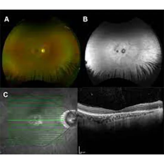
Your optometrist will specify the diagnosis. In the onset, fam history, symptoms like light sensitivity and diminished vision will offer important knowledge for the diagnosis. The retinal examination could appear to be normal. The Farnsworth panel D15 color test, H-R-R tests, Ishihara pseudoisochromatic tests, and City University tests are the four most often used color vision exams in clinics. Making a diagnosis for this disorder requires additional testing, including electroretinograms (ERGs), which measure cone function, optical coherence tomography (OCT), fundus autofluorescence, and visual fields. It is possible to do genetic testing to check for mutations in the most prevalent genes to confirm the diagnosis.
| Test Type | Achromatopsia |
| Includes | Achromatopsia (Pathology Test) |
| Preparation | |
| Reporting | Within 24 hours* |
| Test Price |
₹
|

Early check ups are always better than delayed ones. Safety, precaution & care is depicted from the several health checkups. Here, we present simple & comprehensive health packages for any kind of testing to ensure the early prescribed treatment to safeguard your health.