Scrotal ultrasonography is a technique for visualising the scrotum as well as other organs such as the testicles and epididymis (tubes immediately next to the testicles that collect sperm). The male reproductive organs, the testicles, are located in the scrotum, a flesh-covered pouch between the legs at the base of the penis. Ultrasound imaging of the scrotum creates images of a male's testicles and surrounding tissues by using sound waves. It is the primary method for diagnosing disorders of the testicles, epididymis (tubes directly adjacent to the testicles that collect sperm), and scrotum.
This scan is recommended for a patient who has scrotal pain and oedema.
It aids in the diagnosis of a scrotal cyst or tumour, the size and symmetry of the testicles, the source of testicular pain, and abnormal blood flow in the testicles caused by testicular torsion. It is also advised to ascertain the cause of male infertility.
A testicular ultrasound typically requires no special preparation. There is no need to change your diet, fast, or keep your bladder full before the test.
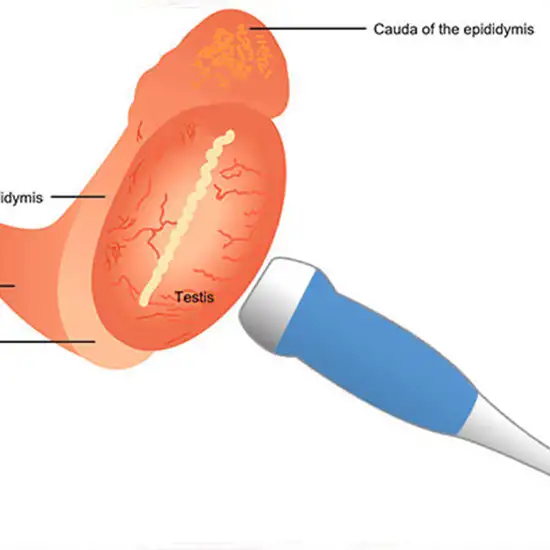
A clear gel is applied to the scrotal sac to aid in the transmission of sound waves. The technologist then moves a handheld probe (the ultrasound transducer) over the scrotum. High-frequency sound waves are produced by the ultrasound machine. These waves create a picture by reflecting off areas of the scrotum.
It probably wouldn't hurt, but you might feel some pressure. The transducer receives sound waves from your skin. A computer screen will display images of your testicles. Following the ultrasound, they will be reviewed by a radiologist.
An Ultrasound Scrotal Test is typically performed in a hospital's radiology department or an outpatient imaging facility. So, for the best services and reliable results, schedule your test at Ganesh Diagnostic in Yamuna Vihar.
