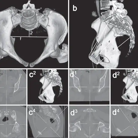
CT Pelvis 3D is an imaging scan that uses 3D reconstruction to visualise the bones, organs, and blood vessels in the pelvic region.
A computed tomography (CT) scan of the pelvis is a type of imaging that uses x-rays to produce cross-sectional images of the area between the hip bones. The pelvic area is the name given to this area of the body.
The bladder, prostate, and other male reproductive organs, female reproductive organs, lymph nodes, small intestine, colon, and pelvic bones are all inside and near the pelvis.
Slices are individual CT images. Images are saved on a computer, displayed on a monitor, or printed on film. By stacking the slices together, three-dimensional models of the body can be created.
You are asked to lie down on a narrow table that slides into the CT scanner's centre.
When you enter the scanner, the x-ray beam rotates around you. The rotating x-ray beams will not be visible.
Because movement causes blurred images, you must remain still during the exam. You might be instructed to hold your breath for brief periods of time.
The scan should take no more than ten minutes.
Doctors advise using this radiology scan to look for:
* Pelvic bone fractures
* Tumours
* Hip dislocations
* Appendicitis
* Infections
* Treatment planning such as biopsy
In pregnancy, CT Pelvis 3D is not performed.
procedure is painless and quick. this procedure is generally well-tolerated, with most patients experiencing either mild pain or no pain during the procedure. There were no patients who experienced excruciating pain.
A CT Pelvis 3D Testis typically performed in a hospital's radiology department or an outpatient imaging facility. So, for the best services and reliable results, schedule your test at Ganesh Diagnostic in Yamuna Vihar.
