Ultrasound imaging of the scrotum creates images of a male's testicles and surrounding tissues by using sound waves. It is the primary method for diagnosing disorders of the testicles, epididymis (tubes directly adjacent to the testicles that collect sperm), and scrotum.
For the Ultrasound of the Scrotum, sound waves provide images of a male's testicles and surrounding tissues. This can be done to look at problems in the testicles and the scrotum.
An ultrasound of the Scrotum is used to analyse the following:
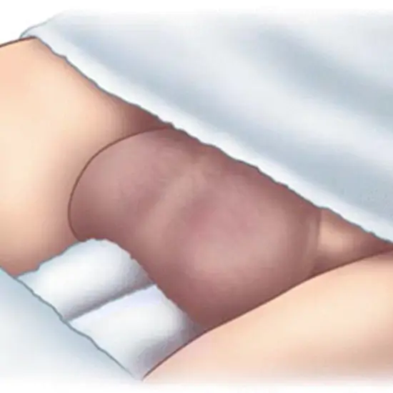
You will lie on your back with your legs spread apart. A cloth should be draped across your thighs under the scrotum, or large strips of sticky tape should be applied to the area. A transparent gel is applied to the scrotal sac, and then a transducer is used to send sound waves to obtain a clear image of the scrotal sac.
A clear gel is applied to the scrotal sac to aid in the transmission of sound waves. The sonographer/physician/radiologist then moves a handheld probe (the ultrasound transducer) over the scrotum. High-frequency sound waves are produced by the ultrasound machine. These waves reflect off the scrotum to form a picture.
Select loose-fitting, comfy clothing. All clothing in the region may need to be removed. For the procedure, you might have to change into a hospital gown.
In most cases, the Ultrasound of Scrotum procedure is painless and can be easily tolerable. You can quickly resume your daily activities after it.
A Scrotum Ultrasound is typically performed in a hospital's radiology department or an outpatient imaging facility. So, for the best services and reliable results, schedule your test at Ganesh Diagnostic in Yamuna Vihar.
