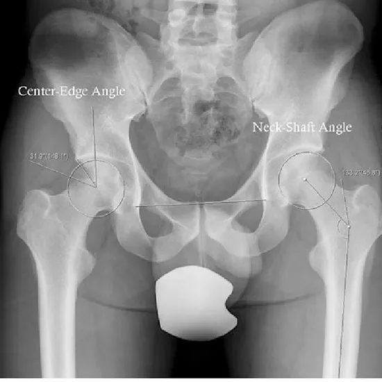
An X-ray of the pelvis is used to see the hip bones, the top part of the thigh bone (called the proximal femur), the hip joint, and the skin and muscles.
• Removal of metallic objects before the procedure is advisable
• Female patient must inform the x-ray technician/ doctor/radiologist about her pregnancy, before undergoing the procedure.
• Patient is lying in a supine position both iliac crests are equidistant to IR or cassette.
• To show an AP view of the proximal femur, the lower limbs are internally rotated 15–25° from the hip (do not attempt this if a fracture is suspected).
