Login

X-RAY ORBITS PA VIEW PROCEDURE

X-RAY ORBITS PA VIEW PROCEDURE

X-RAY ORBITS PA VIEW PROCEDURE

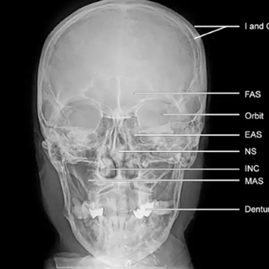
To get a clear orbits (postero-anterior) image of the skull and demonstrate pathology such as skull fractures (orbits) with medial and lateral displacement. Mandible fractures are seen in postero-anterior view. In pediatrics, this perspective is utilized to reduce the equivalent dose to the eye's lens.

This view is useful in assessing:

  • Orbits
  • Internal auditory meatus
  • Skull fracture
  • Paget disease
  • Neoplastic change
  • Frontal bone
  • Ethmoid and frontal sinus
  • Structure seen in dental region
  • Mastoid sinus
  • The sphenoid has larger and smaller wings.

Part positioning

  • The patient is in a seating position in front of a vertical detector.
  • Both forehead and nose are pressed against the IR and touching the detector.
  • At a right angle to the IR, there is an orbito-metal line.
  • The Petrous ridge is below the orbits

Central ray

  • At the level of Nasion, the central ray is pointed perpendicular to the Bucky along the medial sagittal plane.
  • Image must be inclusive of the vertex superiorly and the occipital bone’s base inferiorly.