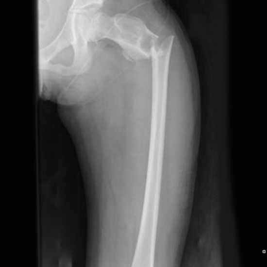
On the radiograph of the proximal femur, the bulk of the shaft, pelvic brim, obturator foramen, acetabulum, ischial spine, femoral head, and femoral neck is visible. The knee joint, the proximal tibia and fibula, the femoral condyles and epicondyles, and the distal two-thirds of the femoral shaft are all visible on a distal femur radiograph. To check for signs of disease or injury in the femur, an x-ray femur AP view is performed. It might be gotten if some clinical signs and symptoms point to a bone injury, or if a femur fracture has been partially imaged on a hip or knee X-ray.
This view is useful in assessing:
• Trauma
• Obvious deformities
• Suspected foreign body
• Inability to weight bear
• Osteomyelitis
• Fracture
These projections are frequently carried out with two images per view due to the image detector's limitations to guarantee the inclusion of both the knee and hip joints.
