Triple Phase Bone Scan
When a fracture cannot be detected on an X-ray, a three-phase bone scan is utilized to make the diagnosis. It is also employed in the diagnosis of osteomyelitis, bone pain, and other disorders affecting the bones.
Indication
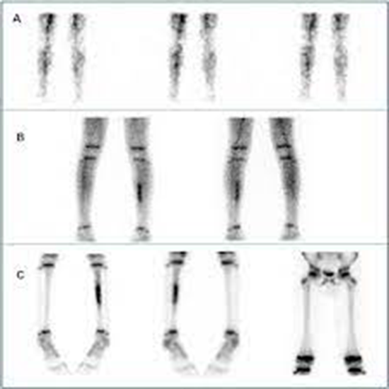
Three-Phase Bone: A bone scan lesion with concomitant hyperperfusion may be present, hence this scan is required to examine blood flow to a particular area. Typical illustrations include:
- Injury (e.g. stress fractures and tendonitis)
- Disease (acute osteomyelitis or septic arthritis)
- CRPS, or Complex Regional Pain Syndrome (formally known as Reflex Sympathetic Dystrophy, RSD)
- Non-cancerous primary tumor
- Primary malignant tumor
- Cold damage (frostbite)
- Tumors of soft tissue (to determine whether adjacent bone is involved)
- Additional skeletal lesions with anticipated corresponding flow anomalies Ossification heterotrophy and avascular necrosis
Preparation
- No preparation is required before the injection; however, the patient should be advised to drink four 8-ounce glasses of liquid after the injection and urged to do so frequently.
- Before imaging, the patient will be asked to empty their bladder. To help with clearance, the patient should be urged to drink a lot of fluids for at least 24 hours following radiopharmaceutical administration.
- For sedated and untrained children, a Foley catheter with a collecting bag should be in place at the time of the delayed scan (second session). *Only required if the pelvis is the area of concern.*
Procedure
- Two visits are required for the test, with a 4-hour gap between each. The initial visit, which lasts typically 20 minutes, starts with the injection of a radioactive isotope into a vein. This injection has no adverse effects.
- The technologist begins taking photographs as soon as the injection is administered. You are free to leave the department following these photos until your subsequent scheduled time.
- Another set of photos will be taken of you during the second visit. Normally, this takes 30 to 60 minutes. Before you leave, a radiologist reviews every image that was taken to decide whether or not more images are necessary.
After Care
The majority of the time, a bone scan is painless and requires no more care afterward. To remove the tracer from your system, you might be instructed to drink a lot of water for the next day or two. Two days following the scan, the radioactivity from the tracers is typically totally gone.
Written by
Ms. SIMRAN KAUSHAL
TECHNOLOGIST(NUCLEAR MEDICINE)