The tissues and organs in the lower abdomen and pelvis can be visualized via ultrasound imaging by using sound waves.
Wear loose-fitting, comfy attire. It might be necessary to take off all of your clothes and valuables in the region being scanned.
For the procedure, you might need to change into a gown.
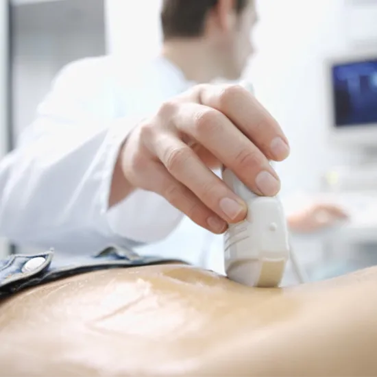
You will be seated on the examination table while the radiologist or sonographer positions the area of your body being examined and applies a water-based gel to it. The sonographer will then apply the transducer to the body and move it back and forth over the desired location until the necessary images are taken. After the imaging is complete, the technologist will remove the clear ultrasonic gel from your skin.
After you've emptied your bladder, the doctor will place the transducer into the vagina. The transducer's tip is smaller than the typical speculum used for Pap tests. The transducer is covered with a protective covering, lubricated with a tiny amount of gel, and inserted into the vagina for two to three inches. To get the best views of the uterus and ovaries, the doctor takes pictures from several perspectives. You will often lie on your back during a transvaginal ultrasound, maybe with your feet in stirrups similar to a gynecologic check-up
The transducer is covered in order to do a transrectal ultrasound. After being lubricated, it is inserted into the rectum.
You typically lay on your side with your knees and hips slightly bent, facing away from the examiner.
After the procedure, the technologist might ask you to change into clothes.
No risks or harmful effects
