MRI BRAIN WITH CERVICAL SPINE PROCEDURE
Magnetic resonance imaging (MRI) of the brain with the cervical spine is an examination used to evaluate the brain, the neck, and the seven cervical spine bones. It can identify a variety of conditions that affect the brain and upper spine, abnormalities like tumors, and problems in the soft tissues within the spinal column, including the spinal cord, nerves, and disks.
Why is MRI Brain with Cervical Done?
- Check for disorders like bruising, swelling, tumors, infections, inflammation, damage from an injury, or a stroke, as well as conditions that affect how the brain formed. Congenital defects or deformities in the spine.
- Inflammation of the spine or tissues near the spine
- Injury/trauma of the spine
- Abnormal/irregular curvature of the spine
- Scoliosis
- Spinal tumor or cancer
- Spinal TB
- Post Spinal surgery evaluation or monitoring.
What is the procedure for MRI Brain with Cervical Spine?
- The consent form explaining the procedure and contrast dye if included in the study is to be signed by the patient or their attendees.
- Any metal objects are to be removed before entering the MRI room.
- Instructions to stay still during the scan and the procedure of the scan are to be explained to the patient.
- The weight of the patient is to be noted.
- If the patient is unstable, sedation can be given.
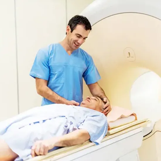
- The patient is positioned supine, head first. Place the head and neck coil on the head of the patient and use the cushions to keep the position intact.
- Provide cushions under the patient legs for comfort.
- Center the laser beam localizer at 2.5cm below the chin making sure the head is in the chin-down position.
Risks
The MRI Brain with Cervical Spine procedure is safe, and the patient can resume normal activities after it is finished.