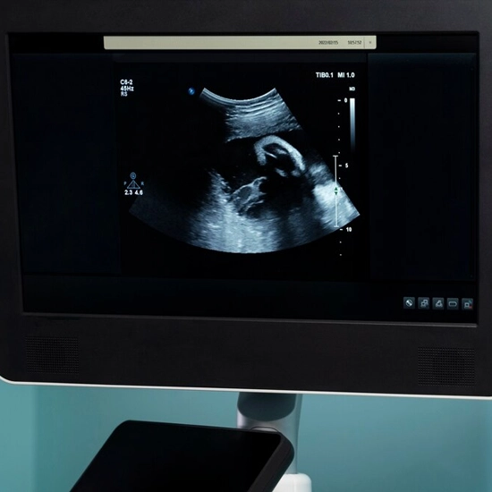
MR SPECTROSCOPY
On the same MRI machine as a standard MRI, MR spectroscopy is performed. A strong magnet, radio waves, and a computer are used in the MRI scan to produce precise images. This scan measures biochemical changes in the brain, especially the presence of tumors. It compares the chemical composition of normal brain tissue with abnormal tumor tissue. MR Spectroscopy is a set of tests that are added to an MRI scan of your brain or spine.
Why is MR Spectroscopy Done?
- Determination of tumor.
- More often done when the cause of the lesion is unclear.
- Precautions for MR Spectroscopy
- Avoid caffeine-containing beverages.
- Since you will be lying still for around 30 minutes, choose comfortable clothing.
- Do not wear metal or jewellery.
Procedure for MR Spectroscopy:
- Your head will be supported by a headrest as you lay on a mobile bed with your arms by your sides.
- To improve the images, contrast dye (gadolinium) may be injected into your arm or administered intravenously.
- A "coil" is a device that will be placed over or around the bodily part being scanned.
- The table will gradually enter the magnetic field once you are in a comfortable position.
- You will experience a prolonged period of muted "thumping" noise as the exam progresses.
Risks
MR spectroscopy is extremely safe. The magnetic field and radio waves used by the device pose no known dangers to human health. Some people are allergic to the contrast agent and may become sensitive to it.
Written By
Ms. Katherine
Education Department