Login

Intravenous Pyelography Procedure

Intravenous Pyelography Procedure

Intravenous Pyelography Procedure

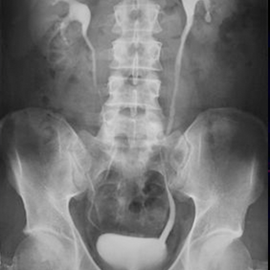
The imaging procedure known as intravenous urography (IVU), commonly referred to as intravenous pyelography (IVP), is used to check the kidney, ureters (the tiny tube that transports urine to the urinary bladder), and the urinary bladder. Monitoring is done of the passage of a contrast dye from the vein through the kidney to the urine bladder.

INDICATION

This view is useful in assessing:

  • Hematuria
  • Dysuria
  • Pyuria
  • Renal colic
  • Bladder pathology
  • Suspected renal trauma
  • Ureter and bladder stone
  • Renal illness
  • Experiencing difficulty peeing
  • Urinary tract infections(UTI)
  • Ureteric duplex
  • Enlarge prostrate
  • Renal, ureteric, and bladder tumor
  • Congenital Anomalies of The Urinary Tract

PATIENT PREPARATION

  • You will likely be instructed to light diet for two days before the procedure and not to eat or drink after midnight on the night before your procedure. You may also be asked to take medication to clear your bowel before the procedure for bowel preparation.
  • Inform your physician of every medicine you are taking
  • Share any recent illnesses or other health concerns with your doctor.
  • The patient’s blood urea and serum creatinine report will be checked before the procedure, if they both are in normal ranges then we can proceed with further examination.
  • Remove the objects containing Metals from the body parts.
  • Notify your radiographic technician in the case of a pregnancy. Unreported radiation may harm the baby

Procedure

  • The patient has to lie in the supine position and take a scout image.
  • If the patient is in good bowel preparation,  then the technician will place a 20G cannula.
  • Then the patient will be given 2ml non-ionic contrast (test dose) on the table to see any allergic reaction through the cannula.
  • 2 mins Nephogram will be taken.
  • 5 mins film will be taken for inflated renal calyx and renal pelvis with abdominal compression band placed on superior iliac crest region.
  • 10 mins. the film will be taken for inflated renal pelvic contrast with the proximal ureter.
  • Then release the compression band with suspended inspiration for 15 mins. The film will be taken for vesico-ureteric junction (VUJ) to the bladder.
  • If VUJ is seen in 15 mins or 20 mins film then the patient needs to take a light diet/ breakfast and wait for full bladder and post-void.
  • If case one renal is not seen then delayed film is taken accordingly nephogram at till time then the patient has to post for 4hrs, 8hrs, and 24hrs film to see their functioning. Or one prone image will be taken for better resolution of the renal region without any blur.