CHEST ULTRASOUND PROCEDURE
A non-invasive diagnostic procedure called a chest ultrasound generates images that are then used to evaluate the organs and structures inside the chest, including the lungs, the region of the chest that contains the heart, aorta, trachea, oesophagus, thymus, and lymph nodes, as well as the space between the lungs and the chest's inner wall. The organs and structures of the chest and blood flow to the organs of the chest may also be evaluated using ultrasound.
Why is Chest Ultrasound Done?
- If your medical professional suspects you have extra fluid in your chest, you could require a chest ultrasound.
- It can be used for looking at the valves in your heart
- Guide a needle to collect a tissue sample
- Examine the movement of your diaphragm
- Check to see whether you have lung fluid accumulation.
Precautions
- Normally, you can continue eating and drinking right up to the exam
- If you believe you might be pregnant or are pregnant, tell your healthcare professional.
- Wear things that are simple to remove. Alternately, dress in a way that the radiologist may access your chest. During the test, a gel is applied to your skin
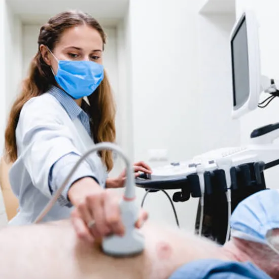
Procedure
- You will lay on your back or your side on the exam table. Alternately, you might sit with your hands clasped behind your neck and your arms lifted.
- A transparent gel will be applied to your skin over the target area by the technologist. The technologist will move the transducer over the area being examined while pressing it firmly against the skin.
- During the exam, you can also be asked to cough or sniff. The technologist will be able to view how your chest's various structures move as a result.
- The technologist will wipe off the gel after the test is finished.
After Care
After a chest ultrasound, no additional care is required.