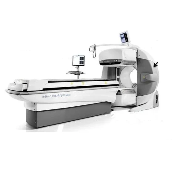
The Single-photon emission computed tomography (also called SPECT or SPET) is a nuclear medicine tomography imaging technique that uses gamma rays.
It is quite similar to a conventional nuclear medicine planar imaging that uses a gamma camera (i.e., Scintigraphy, but this machine is able to provide the true 3D information. This information is usually represented as the cross-sectional slices through the patient, but it can also be freely reformatted or even manipulated as required.
The main advantage of SPET or SPECT is seen as the ability to view the reconstructed image in multiple planes and also to separate the overlapping structures. As much as six-fold increase in image contrast can be obtained with the SPECT machine.