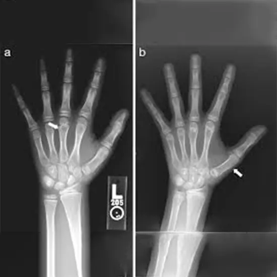
To evaluate a child's skeletal maturity, bone age studies frequently employ X-rays. The body's bones grow and develop at various rates during childhood and adolescence; it is possible to determine a child's...
To evaluate a child's skeletal maturity, bone age studies frequently employ X-rays. The body's bones grow and develop at various rates during childhood and adolescence; it is possible to determine a child's chronological age using their bone growth patterns.
When determining a child's skeletal maturity through bone age studies, X-rays are a crucial tool. The main functions of X-rays in bone age research are listed below:
Assessing bone development
Bone age studies typically involve taking X-rays of the left hand and wrist to evaluate bone development. Numerous bones found in the hand and wrist are a sign of skeletal maturity in general.
Medical practitioners can determine a child's chronological age by looking at the X-ray images and evaluating the bones' degree of growth and maturation.
Certain bones fuse at particular times as the bones in the hand and wrist develop and mature in a predictable order. Healthcare experts can spot any differences or anomalies in bone formation by comparing the X-ray pictures to reference images of bone development.
For instance, it may be a sign of fast skeletal maturation if a child's hand and wrist bones are more developed than would be expected for a child of that age. On the other hand, if the bones are not as formed as you would expect, this may point to a delay in skeletal development.
Predicting growth patterns
Bone age studies can assist forecast how a child's growth patterns may vary over time because the timing and rate of puberty are closely related to skeletal maturity.
For instance, if bone age research reveals that a child's skeletal age is older than their chronological age, this may signal that they will experience puberty earlier than anticipated. On the other hand, a delayed skeletal age might point to a postponement of puberty.
Identifying growth disorders
Analysis of bone ages can be used to spot growth abnormalities in children as well as developmental delays.
For instance, a bone age study can identify children who have delayed skeletal development due to growth hormone insufficiency or other hormonal imbalances.
Similar to how adults may distinguish unique patterns of skeletal development using X-ray imaging, children with diseases that impact bone growth, like Turner syndrome or Down syndrome, may show these patterns as well.
Monitoring treatment progress
Assessing the success of treatment for growth problems or other illnesses that impair skeletal development may occasionally be done using bone age tests.
Healthcare providers can determine whether a child's growth is developing as anticipated and, if required, make adjustments to their treatment plan by comparing X-rays taken at various times in time.
Limits and risks
Although X-rays are typically regarded as safe, they do include exposure to ionizing radiation, which may eventually raise the risk of cancer or other health issues.
Healthcare professionals take precautions to reduce radiation exposure, including using the lowest dose of radiation and shielding other parts of the body that aren't being imaged. Radiation exposure during a bone age study is typically very low.
Furthermore, there are restrictions to bone age studies, and they might not always be reliable. Children who are obese or have certain medical disorders, for instance, may have skeletal development that is different from normal and might be challenging to appropriately measure by X-rays.
X-rays : Exposing the invisible


