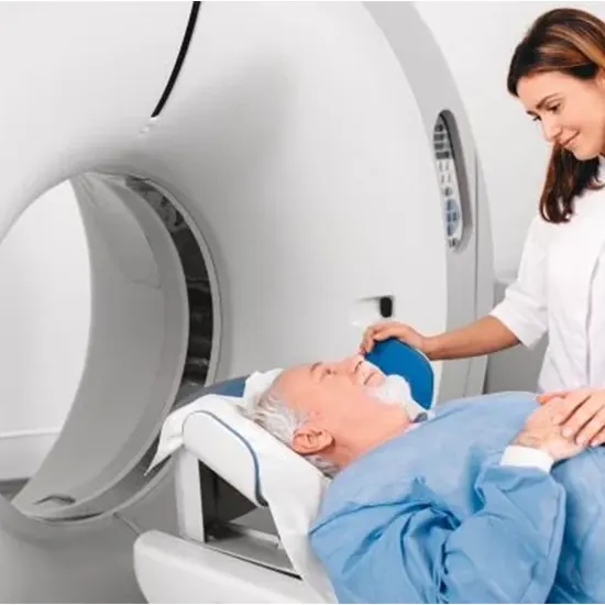
A computed tomography (CT) check, commonly called a CT, may be a radiographic imaging test. The machine was created by physicist Alan MacLeod Cormack and electrical design Godfrey Hounsfield. Their advancement earned them the...
Investigating Inner Structures : A Direct to CT Filters
A computed tomography (CT) check, commonly called a CT, may be a radiographic imaging test. The machine was created by physicist Alan MacLeod Cormack and electrical design Godfrey Hounsfield. Their advancement earned them the 1979 Nobel Prize in Physiology or Medication.
The primary scanner was introduced in 1974. Since then, progresses in innovation and arithmetic have made it conceivable to compute a single outline into his two-dimensional important picture.
A CT filter is fundamentally an x-ray examination in which an arrangement of bars of light are pivoted around a particular portion of the body to make a computer-generated cross-sectional picture.
The advantage is that the pictures don't need to be overlaid as they contain nitty gritty data almost particular regions of the cross area. This can be a huge advantage over customary film. CT checks give great clinicopathologic relationship of suspected disease.
CT looks to progress a doctor's capacity to precisely analyze a patient's illness. Low-dose CT filters have demonstrated valuable in preventive medication and cancer screening. The consideration was initially called CAT Filter. Usually a pivotal CT check where the table moves each time an pivotal picture is taken.
In helical or helical filtering, the table moves persistently as the x-ray source and detector pivot. This essentially abbreviates the examination period and gives speedy comes about in a crisis.
It rapidly supplanted cerebral angiography to identify head injury and brain masses rapidly and dependably. Radiologists acquire CT checks that are deciphered and detailed by prepared radiologists.
Indications
A CT scanner pivots her x-ray tube around the patient's body through a circular structure called a gantry. Computerized data is recorded for each transformation of the machine. The understanding is gradually moved up and down on the sofa to create different cross-sectional pictures.
A cut of her 2D picture of her is created for each turn. The thickness of each consequent picture cut is balanced at the tact of the operator and physician/radiologist, but regularly ranges from 1 to 10 mm. You'll move the gantry to any point to urge the leading cross-sectional picture. Once the desired number of cuts has been procured, the filter is reproduced into a computer picture that can be effortlessly remade and spared.
Pictures are developed utilizing pixels comparing to radio sensitivity and shown utilizing Hounsfield scale units for known tissue densities. for water, -1000 for discussion, +400-2000 for bone. Intravenous iodine can be injected into the circulatory system to portray blood vessels and tumors and identify irresistible forms.
Visualize the gastrointestinal framework utilizing intravenous iodine differentiate or barium differentiate. You'll be able to fasten the images together on your computer to form a 3D picture of him within the region of interest.
CT pictures are taken of cephalad, that's , from the feet to the head. It is vital to note that his current CT scanner shows pictures of the opposite side of the understanding, as the images are produced as seen from the patient's leg. Subsequently, the proper side of the picture is the cleared out side of the persistent.
Tips
CT looks are utilized for numerous clinical indications, depending on the organ examined. CT filters can be used in both inpatient and outpatient clinical settings. In an crisis, genuine ailment can be ruled out. Signs for CT scans are expecting to assist the doctor make a conclusion, limit the differential conclusion, and affirm the physician's doubt.
It can too be utilized for cancer screening, organizing, and follow-up. Its utilize makes a difference to legitimately perform biopsies and give back amid surgical intercessions.
Brain
Tumors, traumatic or unconstrained hematomas, stroke, edema, skull break, calcifications, arteriovenous mutations, hydrocephalus, sinusitis.
Neck
Tumors, generous masses, thyroid knobs, lymphadenopathy.
Chest
Tumor, pneumonia, metastasis, kind masses, pulmonary edema, pleural edema, tuberculosis, pneumonic embolism, traumatic harm to the lungs, esophageal break, ingested outside body, fibrosis.
Guts
Essential tumors, metastases, sore, ascites, cholecystitis, a ruptured appendix, renal calculi, pancreatitis, obstacle, lymphadenopathy, remote body.
Spine
Breaks, degenerative changes, soundness, osteomyelitis, circle pathology.
Bone
Complex bone breaks, dissolved joints, knee, tumors, osteomyelitis.
Gyn
Cyst, fibromas, tumors.
Screening:
Colon and lung cancer.
CT colonography/colonoscopy is utilized to diagnose colon malady and early-stage cancer with great affectability and specificity.
Low-emission CT can be utilized to analyze lung cancer in smokers and previous smokers with a tall smoking history aged between 55 and 80 a long time ancient utilizing a moo radiation dosage.
Biopsy
CT guided to diverse organs for satisfactory tissue extraction.
CT Angiography
Brain, heart, lung, kidney, extremities.
Intraoperative
CT check can be utilized for neuronavigation strategies amid brain biopsy or tumor resection.
Interferometer Factors
CT looks may be considered inconclusive if artifacts obscure the pictures. Interferometer components from metal-based objects such as dental inserts, shrapnel, bullet parts, surgical clips, pacemaker, and body piercings will cause a "flare" known as streak artifacts.
These artifacts in pictures darken fundamental structures and make it troublesome to appropriately visualize and survey dynamic pathology.
To decrease image flickering, metallic artifacts decrease algorithms and normalized metallic artifact lessening progress images and reduce the chance of blunders. In a few cases where past pictures are accessible, a Gaussian dissemination sinogram can be connected to decrease streak artifacts in dental inserts. In any case, this is a constrained examination as past pictures may not be available.
When detailed CT looks are gotten using intravenous differentiate operators, iodine-radiolabeled colors are utilized to identify certain organic markers and clinically pertinent chemical compounds such as troponin, angiotensin-converting protein, or electrolytes such as zinc and iodine.
It can be meddled with assessment. A major downside of CT filters is the poor representation of ligaments, tendons, spinal rope, or intervertebral plates. In such cases, attractive reverberation imaging (MRI) is the test of choice.
Complications
CT scans contain ionizing radiation that damages living tissue. A CT filter can uncover you to 50 to 1,000 times more radiation than a conventional x-ray. They account for most of the radiation from natural/environmental sources within the populace. CT filters make up around 50% of all restorative radiation. It is assessed that there's a 5% chance of developing deadly cancer for each 1.0 mSv of exposure.
A radiation measurement of 100 millisieverts has a 0.5% chance of creating cancer. Generally talking, for every 1000 CT filters of a pediatric quiet she has, he develops one deadly cancer. Utilizing nuclear bomb data, a pediatric head CT check encompasses a lifetime chance of 1 in 10,000 for leukemia and 2,000 to 10,000 for brain tumors.
This radiation presentation is especially basic in pediatric patients due to the helplessness of creating organs and aggregate lifetime presentation in case gotten some time recently the age of ten. Exposures ought to be restricted concurring to the ALARA guideline (as moo as sensibly achievable).
They should be implemented when the benefits far exceed the dangers. Radiation measurements for CT looks run from 1.0 mSv to 27.0 mSv. 1 millisievert = 1 milligray. The natural/environmental introduction is roughly 3.0 mSv per year. A grown-up stomach CT uncovered the patient to 10 mSv.
In any case, the presentation measurements for neonatal abdominal CT is 20 mSv.
Differentiate operators can cause allergic responses, which are ordinarily mellow and incorporate a bothersome hastiness. In any case, genuine responses such as bronchospasm and anaphylactic responses can happen.
The chance of a lethal response is about 1 in 100,000. On the off chance that you are allergic to iodine, you may have to be regulate steroids to decrease the side impacts of contrast media organization. Iodinated differentiate renal failure can occur in 2% to 7% of patients, with patients with pre-existing renal disease at expanded chance.
In the event that differentiate nephropathy is serious, dialysis may be required to expel the difference. In non-severe cases, drinking sufficient liquids some time recently infusing the differentiate medium will offer assistance it pass from the body.
Quiet security and instruction
The evaluated radiation measurements gotten in a single filter compares to characteristic human introduction to the environment over months to a long time. If the patient is pregnant, the radiation measurements is little and as a rule does not influence the baby.
In any case, this investigation will as it were conducted on the off chance that there is a fabric advantage. A winding or spiral CT scan reduces the radiation measurements compared to a sequential CT check.
Low-emission CT looks have the advantage of minimizing radiation presentation and early discovery of cancer in smokers. CT checks are perfect for injury since they can identify organ harm rapidly and reliably. A venous differentiation upgrades the vascular structure and can recognize the cause of a subarachnoid hemorrhage or unconstrained hematoma.
In the case of gunshot wounds, CT scans give points of interest of the harm, while MRI cannot do this due to the metallic nature of discharge wounds. CT filters have several advantages over attractive reverberation imaging.
CT scans do not affect device settings and are safe for patients with pacemakers or programmable pumps or shunts. Patients with claustrophobia ordinarily cannot experience MRI looks. In any case, CT looks are speedier, calmer, and more comfortable.


