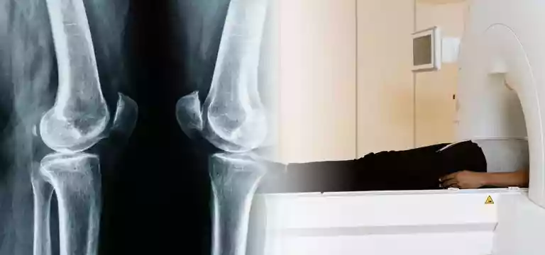
MRI of the left leg is a scan that focuses on the evaluation of left leg pathologies. It is important to note that the left leg is a complex anatomical structure consisting of bones, muscles, tendons, ligaments, and blood...
Introduction
MRI of the left leg is a scan that focuses on the evaluation of left leg pathologies. It is important to note that the left leg is a complex anatomical structure consisting of bones, muscles, tendons, ligaments, and blood vessels. As a result, it is vulnerable to a wide range of conditions and injuries. So, as a powerful imaging modality, an MRI of the left leg plays a significant role in diagnosing and assessing these pathologies with the help of detailed and high-resolution images of the leg.
In this article, we will discover the various left leg pathologies that an MRI can evaluate, such as fractures, sprains, strains, ligament tears, muscle tears, and tumors. We will also go into the specific imaging techniques and protocols used in MRI examinations of the left leg.
It is important to research and know about the role of an MRI in left leg pathologies, assisting healthcare professionals in providing enhance patient care.
What does a left leg MRI entail?
When it comes to the field of orthopedics and musculoskeletal imaging, an MRI can be an invaluable imaging technique that has upended medical diagnostics. So, in evaluating left leg pathologies, MRI, with its capability to churn out detailed and multi-planar images, has helped healthcare professionals to accurately visualize and assess the intricate structures of the left leg. It provides accurate images of bones, muscles, tendons, ligaments, and blood vessels, immensely helping doctors to treat patients.
It helps in assessing various conditions such as fractures, ligamentous injuries, muscle tears, tendonitis, tumors, and other abnormalities of the soft tissue. By generating high-resolution images, MRI helps zero in on the precise location, extent, and severity of these pathologies, aiding accurate diagnosis and improving treatment planning.
Moreover, MRI has the advantage of being non-invasiveness and there is no ionizing radiation, which makes it a safe and effective imaging technique in evaluating left leg pathologies.
Overall, MRI’s importance in imaging the left leg is unparalleled, providing great assistance to the doctors involved in offering targeted and individualized care to patients and improving their quality of life.
Left leg MRI: The process and requirements
What is a comprehensive left leg MRI examination all about? It is referred to the process of capturing detailed images of the key components that constitute the complex anatomy of the left leg.
It is based on the process of involving several essential sequences and imaging parameters. To study the bony structures, T1-weighted and T2-weighted sequences are commonly employed. They can provide near-perfect images of the bones, including the femur, tibia, fibula, and foot bones. For analyzing soft tissues such as muscles, tendons, and ligaments, proton density-weighted or fat-suppressed sequences are utilized. They enhance the visibility of these structures and facilitate identifying any abnormalities like strains, tears, or inflammation.
A contrast-enhanced MRA (Magnetic Resonance Angiography) sequence helps in examining blood vessels, thereby helping in studying arterial and venous structures. It unravels conditions like deep vein thrombosis or peripheral arterial disease.
It's paramount to optimize imaging parameters such as field of view (FOV), slice thickness, and imaging time to get improved quality images and diagnostic accuracy. In certain cases, the radiologist may also use multiplanar reconstructions and 3D rendering techniques. It helps them in grasping deep insights into the left leg anatomy.
Hence, taking these key components into account and optimizing imaging parameters, doctors can conduct a comprehensive left leg MRI examination. Equipped with this knowledge from the MRI scan, doctors are capable of deriving an accurate diagnosis and guiding appropriate treatment interventions for patients.
Musculoskeletal Injuries: Diagnosis and Management
It is a complex process to diagnose and manage musculoskeletal injuries, which involves a comprehensive approach. It is with a thorough clinical evaluation its diagnosis begins. The doctors try to get a detailed history and physical examination as a part of the process.
They employ many imaging techniques such as X-rays, Magnetic Resonance Imaging (MRI), and Computed Tomography (CT) scans to derive a detailed assessment of the injury.
These modalities of imaging help in analyzing fractures, dislocations, ligamentous tears, muscle strains, and other soft tissue injuries.
Once healthcare professionals achieve a correct diagnosis, they focus on the management of musculoskeletal injuries aiming to relieve pain, promote healing, and restore function.
Physicians may resort to conservative measures such as rest, physical therapy, pain management techniques, and the use of orthotics or braces for treating patients.
In some severe cases, they may opt for surgical intervention to repair or reconstruct damaged structures. It is important to note that rehabilitation plays a key role in the recovery process. Its main aim is to regain strength, flexibility, and mobility.
An individual would require close collaboration between healthcare professionals, including orthopedic specialists, physical therapists, and sports medicine experts, to derive the optimum benefit from this treatment plan. Doctors manage to provide optimal recovery, helping patients regain functionality with the help of an accurate diagnosis and proper management of musculoskeletal injuries.
Challenges with Left leg MRI
Apart from all the advantages of a left leg MRI scan, there may some challenges for some patients. It may be related to patient positioning, motion artifacts, and claustrophobia that need to be effectively addressed for a successful imaging session.
In any MRI examination, patient positioning is critical to obtain accurate and high-quality images. It is important to use immobilization devices, like foam pads or straps, so that the leg is stabilized from motion artifacts caused by involuntary movements.
It is also important to establish constant communication with the patient and make them understand the importance of remaining still during the scan. There are techniques to achieve this such as breath-holding instructions or triggering the scan during specific phases of respiration. It will help to further minimize motion artifacts.
Claustrophobia is another key impediment the healthcare personnel have to address during MRI examinations. Patients who suffer from this need some assurance from the technicians during the scan.
Left leg MRI Price
To ascertain the price of a left leg MRI we have to consider several factors that may have a bearing on the overall cost. The left leg MRI price can vary depending on the region, the healthcare facility or imaging center, and any extra services required.
Another key area to consider is the insurance coverage a patient has. The healthcare providers may also have a role to play in determining the final price of an MRI. So, talk to your insurance company for any coverage you may have before going for the left leg MRI price. Ensure what your entire insurance plan covers.
Find out by discussing with your healthcare facility any potential hidden costs, such as radiologist interpretation fees or facility fees. It will give you a clear understanding of the total cost.
Conclusion
So, we have seen that a left leg MRI is a specialized imaging procedure that focuses on capturing detailed images of the structures within the left leg. It is a non-invasive and painless diagnostic tool that utilizes powerful magnetic fields and radio waves to generate high-resolution images of the bones, muscles, tendons, ligaments, and blood vessels in the left leg.
Healthcare professionals use this technique more commonly to diagnose and evaluate various conditions such as fractures, sprains, ligament tears, muscle strains, tumors, and other abnormalities affecting the left leg.
The images obtained from a left leg MRI lends invaluable information to the doctors for accurate diagnosis, treatment planning, and monitoring of the leg's health. By looking at the internal structures through a left leg MRI doctors can identify the root cause of symptoms, take informed decisions on interventions, and facilitate the highest patient care.
FAQ
Why do doctors recommend a left leg MRI?
Doctors usually recommend a left leg MRI to evaluate and diagnose various conditions or injuries affecting the bones, muscles, tendons, ligaments, or blood vessels in a patient’s left leg.
What can a patient expect during a left leg MRI?
During a left leg MRI, the patient will be asked to lie down on a movable table that slides into a large cylindrical machine. One has to remain still during the process. Then the machine will begin to take images of the left leg. The machine also produces loud noises, but ear protection is generally provided.
Is a left leg MRI painful?
No, a left leg MRI is not painful. But some people may experience mild discomfort due to lying down for an extended period. If a patient has such concerns, he or she should discuss them with the technicians and doctors about it so that they can provide some available solutions for this. Overall, it is an easy process most of the time.


